
Page 768 - Cardiac Nursing
P. 768
744 P AR T IV / Pathophysiology and Management of Heart Disease
treatment of native and recurrent coarctation, particularly in
teenagers and young adults who have achieved full growth. 29
Patients with coarctation of the aorta are prone to arterial hy-
pertension and coronary heart disease, due to arterial wall stiff-
ness and alterations in vascular reactivity. 30–33 Functional data
and histological findings suggest a systemic vascular disease of the
prestenotic arteries, even after successful surgical repair. 30,34 In
several studies, CHD was found to be the most common cause of
late death in patients with coarctation of the aorta. 35–38 Therefore,
aggressive risk factor management for prevention of general ac-
quired heart disease should be undertaken. ACE inhibitors and
b-blockers are particularly useful in the management of these pa-
39
tients. Other long-term residual effects include restenosis of the
previously treated area and aneurysms of the ascending aorta.
Follow-up for patients with coarctation of the aorta is required
to monitor for late complications such as hypertension and
restenosis. Such follow-up and subsequent treatment should be
scheduled every 1 to 2 years 26 in an adult congenital heart disease
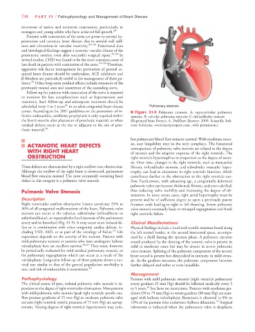
center. According to the 2007 guidelines on the prevention of in- n Figure 31-9 Pulmonic stenosis. A: supravalvular pulmonic
fective endocarditis, antibiotic prophylaxis is only required within stenosis; B: valvular pulmonic stenosis; C: infundibular stenosis
the first 6 months after placement of prosthetic material, or when (Reprinted from Everett, A. PedHeart Resource. 2009. Scientific Soft-
residual defects occur at the site or adjacent to the site of pros- ware Solutions. www.heartpassport.com., with permission.)
thetic material. 20
but pulmonary blood flow remains normal. With moderate steno-
ACYANOTIC HEART DEFECTS sis, easy fatigability may be the only complaint. The functional
WITH RIGHT HEART consequences of pulmonic valve stenosis are related to the degree
of stenosis and the adaptive response of the right ventricle. The
OBSTRUCTION right ventricle hypertrophies in proportion to the degree of steno-
sis. Over time, changes in the right ventricle, such as myocardial
These defects are characterized by a right outflow tract obstruction. fibrosis, infundibular stenosis, and subvalvular muscular hyper-
Although the outflow of the right heart is obstructed, pulmonary trophy, can lead to alterations in right ventricle function, which
blood flow remains normal. The most commonly occurring heart contributes further to the obstruction to the right ventricle out-
defect in this category is a pulmonic valve stenosis. flow. Furthermore, with advancing age, a congenitally deformed
pulmonic valve can become thickened, fibrotic, and even calcified,
Pulmonic Valve Stenosis thus reducing valve mobility and increasing the degree of ob-
struction. In more severe cases, right atrial hypertrophy must be
Description present and be of sufficient degree to open a previously patent
Right ventricular outflow obstructive lesions constitute 25% to foramen ovale leading to right to left shunting. Severe pulmonic
30% of all congenital malformations of the heart. Pulmonic valve valve stenosis eventually leads to tricuspid regurgitation and frank
stenosis can occur at the valvular, subvalvular (infundibular or right ventricle failure.
subinfundibular), or supravalvular level (stenosis of the pulmonary
artery and its branches) (Fig. 31-9). It may occur as an isolated de- Clinical Manifestations
fect or in combination with other congenital cardiac defects, in- Physical findings include a loud mid-systolic murmur heard along
cluding VSD, ASD, or as part of the tetralogy of Fallot. 13 Life the left sternal border, at the second intercostal space, accompa-
expectancy depends on the severity of the stenosis. Patients with nied by a thrill during the ejection phase. A pulmonic ejection
mild pulmonary stenosis or patients who have undergone balloon sound produced by the doming of the stenotic valve is present in
valvuloplasty have an excellent survival. 40,41 They must, however, mild to moderate cases but may be absent in severe pulmonic
be periodically evaluated for mild residual pulmonary stenosis, or valve stenosis. Splitting of the pulmonic component of the second
for pulmonary regurgitation which can occur as a result of the heart sound is present but diminished in intensity in mild steno-
valvuloplasty. Long-term follow-up of these patients shows a sur- sis. As the gradient increases, the pulmonic component becomes
vival rate similar to that of the general population; morbidity is further delayed and softer or even inaudible.
rare, and risk of endocarditis is nonexistent. 42
Management
Pathophysiology Patients with mild pulmonic stenosis (right ventricle pulmonary
The clinical course of pure, isolated pulmonic valve stenosis is de- artery gradient 25 mm Hg) should be followed medically every 3
pendent on the degree of right ventricular obstruction. Most patients to 5 years, 26 but have no restrictions. Patients with moderate gra-
with mild pulmonic valve stenosis (peak right ventricle systolic out- dients (50 to 79 mm Hg) or severe gradient (80 mm Hg) are man-
flow pressure gradients of 25 mm Hg) to moderate pulmonic valve aged with balloon valvuloplasty. Restenosis is observed in 8% to
43
stenosis (right ventricle systolic pressures of 75 mm Hg) are asymp- 10% of the patients who underwent balloon dilatation. Surgical
tomatic. Varying degrees of right ventricle hypertension may exist, valvotomy is indicated when the pulmonary valve is dysplastic