
Page 112 - untitled
P. 112
AAAC48 21/5/05 10:52 AM Page 111
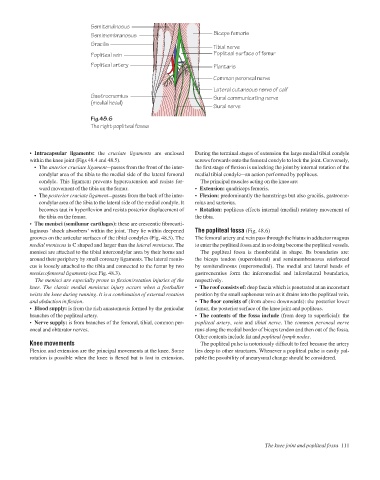
Semitendinosus
Semimembranosus
Gracilis Biceps femoris
Tibial nerve
Popliteal vein Popliteal surface of femur
Popliteal artery Plantaris
Common peroneal nerve
Lateral cutaneous nerve of calf
Gastrocnemius Sural communicating nerve
(medial head)
Sural nerve
Fig.48.6
The right popliteal fossa
• Intracapsular ligaments: the cruciate ligaments are enclosed During the terminal stages of extension the large medial tibial condyle
within the knee joint (Figs 48.4 and 48.5). screws forwards onto the femoral condyle to lock the joint. Conversely,
• The anterior cruciate ligamentapasses from the front of the inter- the first stage of flexion is unlocking the joint by internal rotation of the
condylar area of the tibia to the medial side of the lateral femoral medial tibial condyleaan action performed by popliteus.
condyle. This ligament prevents hyperextension and resists for- The principal muscles acting on the knee are:
ward movement of the tibia on the femur. • Extension: quadriceps femoris.
• The posterior cruciate ligamentapasses from the back of the inter- • Flexion: predominantly the hamstrings but also gracilis, gastrocne-
condylar area of the tibia to the lateral side of the medial condyle. It mius and sartorius.
becomes taut in hyperflexion and resists posterior displacement of • Rotation: popliteus effects internal (medial) rotatory movement of
the tibia on the femur. the tibia.
• The menisci (semilunar cartilages): these are crescentic fibrocarti-
laginous ‘shock absorbers’ within the joint. They lie within deepened The popliteal fossa (Fig. 48.6)
grooves on the articular surfaces of the tibial condyles (Fig. 48.5). The The femoral artery and vein pass through the hiatus in adductor magnus
medial meniscus is C shaped and larger than the lateral meniscus. The to enter the popliteal fossa and in so doing become the popliteal vessels.
menisci are attached to the tibial intercondylar area by their horns and The popliteal fossa is rhomboidal in shape. Its boundaries are:
around their periphery by small coronary ligaments. The lateral menis- the biceps tendon (superolateral) and semimembranosus reinforced
cus is loosely attached to the tibia and connected to the femur by two by semitendinosus (superomedial). The medial and lateral heads of
meniscofemoral ligaments (see Fig. 48.3). gastrocnemius form the inferomedial and inferolateral boundaries,
The menisci are especially prone to flexion/rotation injuries of the respectively.
knee. The classic medial meniscus injury occurs when a footballer • The roof consists of: deep fascia which is penetrated at an inconstant
twists the knee during running. It is a combination of external rotation position by the small saphenous vein as it drains into the popliteal vein.
and abduction in flexion. • The floor consists of (from above downwards): the posterior lower
• Blood supply: is from the rich anastomosis formed by the genicular femur, the posterior surface of the knee joint and popliteus.
branches of the popliteal artery. • The contents of the fossa include (from deep to superficial): the
• Nerve supply: is from branches of the femoral, tibial, common per- popliteal artery, vein and tibial nerve. The common peroneal nerve
oneal and obturator nerves. runs along the medial border of biceps tendon and then out of the fossa.
Other contents include fat and popliteal lymph nodes.
Knee movements The popliteal pulse is notoriously difficult to feel because the artery
Flexion and extension are the principal movements at the knee. Some lies deep to other structures. Whenever a popliteal pulse is easily pal-
rotation is possible when the knee is flexed but is lost in extension. pable the possibility of aneurysmal change should be considered.
The knee joint and popliteal fossa 111