
Page 111 - untitled
P. 111
AAAC48 21/5/05 10:52 AM Page 110
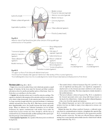
Medial condyle
Posterior cruciate ligament
Lateral condyle Anterior cruciate ligament
Medial meniscus
Fibular collateral ligament Coronary ligament
Fat
Ligamentum patellae
Tibial collateral ligament
Patella (turned back)
Fig.48.4
Anterior view of the flexed knee joint after division of the quadriceps
and retraction of the patella
Deep infrapatellar
bursa
Transverse ligament Cut edge of
capsular ligament
Anterior cruciate
ligament Lateral meniscus
Tibial collateral Fibular collateral
ligament ligament
Medial meniscus Posterior menisco-
femoral ligament Anterior Posterior
Tendon of popliteus cruciate ligament cruciate ligament
Posterior cruciate ligament
Fig.48.5
The upper surface of the tibia and related structures.
The dotted line indicates the synovial membrane in the vicinity of the cruciate ligaments.
The small diagrams show how the cruciate ligaments resist forward and backward displacement of the femur
The knee joint (Figs 48.1–48.5) • The medial (tibial) collateral ligament (Figs 48.1 and 48.3)acon-
• Type: it is a synovial modified hinge joint which also permits a small sists of superficial and deep parts. The superficial component is
degree of rotation. In the knee joint the femoral and tibial condyles attached above to the femoral epicondyle and below to the subcuta-
articulate as does the patella and patellar surface of the femur. Note: the neous surface of the tibia. The deep component is firmly attached to
fibula does not contribute to the knee joint. the medial meniscus.
• Capsule: the articular surfaces are covered by articular cartilage. The • The lateral (fibular) collateral ligament (Fig. 48.2)ais attached to
capsule is attached to the margins of the articular surfaces except anter- the femoral epicondyle above and, along with biceps femoris, to
iorly where it dips downwards. In the anterior part of the capsule there the head of the fibula below. Unlike the medial collateral ligament
is a large opening through which the synovial membrane is continuous it lies away from the capsule and meniscus.
with the suprapatellar bursa (Fig. 48.2). This bursa extends superiorly The collateral ligaments are taut in full extension and it is in this
three fingerbreadths above the patella between the femur and quad- position that they are liable to injury when subjected to extreme val-
riceps. When an effusion has collected fluctuation can be elicited. gus/varus strain.
Posteriorly the capsule communicates with another bursa under the Behind the knee the oblique popliteal ligament, a reflected exten-
medial head of gastrocnemius and often, through it, with the bursa of sion from the semimembranosus tendon, strengthens the capsule (Fig.
semimembranosus. Posterolaterally another opening in the capsule per- 48.3). Anteriorly the capsule is reinforced by the ligamentum patellae
mits the passage of the tendon of popliteus. and the patellar retinacula. The latter are reflected fibrous expansions
• Extracapsular ligaments: the capsule of the knee joint is reinforced arising from vastus lateralis and medialis muscles which blend with the
by ligaments. capsule anteriorly (Fig. 48.1).
110 Lower limb