
Page 150 - untitled
P. 150
AAAC67 21/5/05 11:01 AM Page 149
Masseter
Facial nerve
Parotid duct
Parotid
Mandible
Sternocleidomastoid
Medial pterygoid
Mastoid process
Retromandibular vein
Posterior belly of digastric
Maxillary artery
Carotid sheath
Wall of pharynx
External carotid artery
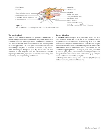
Fig.67.2 Styloid process and 3 'stylo-' muscles
A horizontal section through the parotid to show its relations
The parotid gland Nerves of the face
Situated mainly behind the mandible but spills over it onto the face. It • The facial nerve: having left the stylomastoid foramen, the facial
extends deeply to come into contact with the pharynx and posteriorly it nerve enters the parotid and divides into frontal, zygomatic, buccal,
is moulded around the mastoid process and sternomastoid. The parotid marginal mandibular and cervical branches (Fig. 67.3), with some
duct extends forwards across masseter to enter the mouth opposite intercommunicating branches between them. Note that the marginal
the second upper molar. The whole gland is enclosed in dense fascia so mandibular branch lies below the mandible for part of its course so that
that swelling of the gland, as in mumps for instance, is very painful. submandibular incisions are made well below the mandible. The cer-
Three structures pass through the gland (Fig. 67.2). These are, from vical branch supplies platysma. Lesions of the facial nerve, for example
superficial to deep: the facial nerve, the retromandibular vein (the by tumours of the parotid, cause unilateral drooping of the face with
beginning of the external jugular) and the external carotid artery, with loss of the normal skin creases, and it can be shown up by asking the
its maxillary and superficial temporal branches. patient to whistle or close up his eyes tightly.
• The trigeminal nerve: sensory to the whole face (Fig. 67.3) except
for the area over the parotid (see Chapter 57).
The face and scalp 149