
Page 151 - untitled
P. 151
AAAC67 21/5/05 11:01 AM Page 150
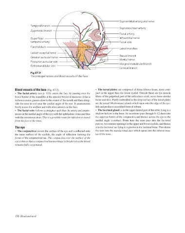
Supraorbital artery and nerve
Temporal branch
Supratrochlear artery
Zygomatic branch
Facial artery
Superficial Infraorbital nerve
temporal artery Facial vein
Parotid duct
Labial branches
Lesser occipital nerve
Buccal branch
Greater auricular nerve
Mental nerve
Posterior auricular vein
Marginal mandibular branch
Retromandibular vein
Cervical branch
Fig.67.3
The principal nerves and blood vessels of the face
Blood vessels of the face (Fig. 67.3) • The tarsal plates: are composed of dense fibrous tissue, more com-
• The facial artery (see p. 133): enters the face by passing over the pact in the upper than the lower eyelid. Outside these are the muscle
lower border of the mandible at the anterior border of masseter. It has a fibres of the palpebral part of the orbicularis oculi, some loose areolar
tortuous course, passes close to the corner of the mouth and then along- tissue and skin. Partly embedded in the deep surface of the tarsal plates
side the nose to end near the medial angle of the eye. It anastomoses are the tarsal (Meibomian) glands which open onto the edge of the eye-
freely across the midline and with other arteries on the face. lids and produce a modified form of sebum.
• The facial vein: follows a straighter path than the artery and anasto- • The lacrimal gland: is in the upper lateral part of the orbit, lying in a
moses at the medial angle of the eye with the ophthalmic veins and thus shallow hollow in the bone. Its secretions pass through 9–12 ducts into
with the cavernous sinus. This is a possible route for infection to travel the superior fornix of the conjunctiva and thence across the eye to the
from the face to the sinus. medial angle (canthus). From here the tears pass into the lacrimal
puncta, two minute openings in the upper and lower eyelids, and thence
The eye into the lacrimal sac lying in a groove in the lacrimal bone. This drains
• The conjunctiva: covers the surface of the eye and is reflected onto the tears into the nasolacrimal duct which opens into the inferior mea-
the inner surface of the eyelids, the angle of reflection forming the tus of the nose.
fornix of the conjunctival sac. The conjunctiva over the surface of the
eye is thin so that a conjunctival haemorrhage is bright red as the blood
remains fully oxygenated.
150 Head and neck