
Page 156 - untitled
P. 156
AAAC69 21/5/05 11:00 AM Page 155
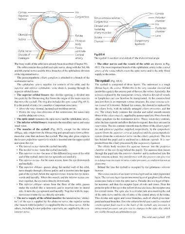
Iris
Cornea
Aqueous
Conjunctiva
Anterior
Sinus venosus sclerae
humour
chamber
Conjunctiva Cornea
Iris
Circular and
Posterior
radiating
chamber
ciliary muscles
Choroid Retina Lens
Sclera
Sclera
Vitreous Choroid Suspensory
body ligament
Fovea
Optic nerve centralis
Sheath of Central Fig.69.4
meninges artery The eyeball in section and detail of the iridocorneal angle
The bony walls of the orbit have already been described (Chapter 55). • The other nerves and the vessels of the orbit are shown in Fig.
The orbit contains the eyeball and optic nerve, along with the 3rd, 4th 69.3. The most important branch of the ophthalmic artery is the central
and 6th cranial nerves and the three branches of the ophthalmic division artery of the retina which enters the optic nerve and is the only blood
of the trigeminal nerve. supply to the retina.
The parasympathetic ciliary ganglion is attached to a branch of the
oculomotor nerve. The eyeball (Fig. 69.4)
The ophthalmic artery supplies the contents of the orbit, and the The eyeball is composed of three layers. The outermost is a tough
superior and inferior ophthalmic veins drain it, passing through the fibrous layer, the sclera. Within this is the very vascular choroid and
superior orbital fissure. inside this again is the sensory part of the eye, the retina. Anteriorly, the
• The superior orbital fissure: this slit-like opening is divided into sclera is replaced by the transparent cornea, which is devoid of vessels
two parts by the fibrous ring that forms the origin of the main muscles or lymphatics and can therefore be transplanted. At the corneoscleral
that move the eyeball. The ring also includes the optic canal (Fig. 69.1). junction there is an important venous structure, the sinus venosus scle-
It is the portal of entry for a number of important structures: rae (canal of Schlemm). Behind the cornea, the choroid is replaced by
• Above the ringafrontal, lacrimal and trochlear nerves. the ciliary body, with its radially arranged ciliary processes, and the
• Within the ringatwo divisions of the oculomotor, the nasociliary iris. The ciliary body contains the circular and radial smooth muscle
and the abducent nerves. fibres of the ciliary muscle, supplied by parasympathetic fibres from the
• The optic canal: transmits the optic nerve and the ophthalmic artery. ciliary ganglion via the oculomotor nerve. These, when they contract,
• The inferior orbital fissure: transmits the maxillary nerve and some relax the lens capsule and allow the lens to expand; thus they are used in
small veins. near vision. The iris contains smooth muscle fibres of the dilator pupil-
• The muscles of the eyeball (Fig. 69.2), except for the inferior lae and sphincter pupillae, supplied, respectively, by the sympathetic
oblique, take origin from the fibrous ring and spread out to form a fibro- system (from the superior cervical ganglion) and the parasympathetic
muscular cone that encloses the eyeball. The ring also gives origin to system (from the oculomotor nerve via the ciliary ganglion). The lens
the levator palpebrae superioris which is inserted into the upper eyelid lies behind the pupil and is enclosed in a delicate capsule. It is sus-
and opens the eye. pended from the ciliary processes by the suspensory ligament.
• The lateral rectusaturns the eyeball laterally. The ciliary body secretes the aqueous humour into the posterior
• The medial rectusaturns the eyeball medially. chamber of the eye (lying behind the pupil). The aqueous then passes
• The superior rectusabecause of the different long axes of the orbit through the pupil into the anterior chamber and is reabsorbed into the
and of the eyeball, turns the eye upwards and medially. sinus venosus sclerae. Any interference with this process can give rise
• The inferior rectusafor the same reason, turns the eye downwards to a dangerous increase in intra-ocular pressure, a condition known as
and medially. glaucoma.
• The superior obliqueapasses along the medial wall of the orbit, Behind the lens the eyeball is occupied by the gelatinous vitreous
turns sharply through a fibrous pulley and is inserted into the upper humour.
part of the eyeball, below the superior rectus. It turns the eye down- The retina consists of an inner nervous layer and an outer pigmented
wards and laterally. When this muscle and the inferior rectus con- layer. The nervous layer has an innermost layer of ganglion cells whose
tract together, the eye turns directly downwards. axons pass back to form the optic nerve. Outside this is a layer of bipo-
• The inferior obliqueaarises from the floor of the orbit, passes lar neurones and then the receptor layer of rods and cones. Near the
under the eyeball like a hammock and is inserted into its lateral posterior pole of the eye is the yellowish macula lutea, the receptor area
side. It turns the eye upwards and laterally. Together with the supe- for central vision. The optic disc is a circular pale area marking the end
rior rectus it turns the eye directly upwards. of the optic nerve and the site of entry of the central artery of the retina.
• The nerve supply of the orbital muscles: the lateral rectus (‘abduc- This divides into upper and lower branches, each of which gives tem-
tor’) of the eye is supplied by the abducent nerve, the superior rectus poral and nasal branches. Since the subarachnoid space and its contained
(the ‘muscle with the pulley’) is supplied by the trochlear nerve. All the cerebrospinal fluid reach to the back of the eyeball, any increase in
others, including levator palpebrae superioris, are supplied by the ocu- intracranial pressure can give rise to changes in the optic disc which
lomotor nerve. are visible through an ophthalmoscope.
The orbit and eyeball 155