
Page 188 - Color Atlas Of Pathophysiology (S Silbernagl Et Al, Thieme 2000)
P. 188
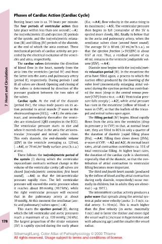
Phases of Cardiac Action (Cardiac Cycle)
       Resting heart rate is ca. 70 beats per minute.  (IIa; → A4), flow velocity in the aorta rising to
       The four periods of ventricular action thus  its maximum (→ A5). The ventricular pressure
       take place within less than one second (→ A):  then begins to fall (remainder of the SV is
       the iso(volu)metric (I) and ejection (II) periods  ejected more slowly, IIb), finally to below that
       of the systole, and the iso(volu)metric relaxa-  in the aorta and pulmonary artery, when the
       tion (III) and filling (IV) periods of the diastole,  semilunar valves close (second heart sound).
                                                             2
       at the end of which the atria contract. These  The average SV is 80 mL (47 mL/m b.s.a.), so
       mechanical periods of cardiac activity are pre-  that the ejection fraction (= SV/EDV) is about
       ceded by the electrical excitation of the ventri-  0.67 at rest. Thus, a residual volume of ca.
       cles and atria, respectively.   40 mL remains in the ventricle (endsystolic vol-
         The cardiac valves determine the direction  ume [ESV]; → A4).
                                        Diastole now begins with the iso(volu)met-
       of blood flow in the heart, namely from the
    Heart and Circulation  the latter into the aorta and pulmonary artery  atria have filled again, a process to which the
                                       ric relaxation period (III). In the meantime the
       atria into the ventricles (period IV) and from
       (period II), respectively. During periods I and
                                       suction effect produced by the lowering of the
                                       valve level (momentarily enlarging atrial vol-
       III all valves are closed. Opening and closing of
                                       ume) during the ejection period has contribut-
       the valves is determined by direction of the
                                       ed the most (drop in the central venous pres-
       pressure gradient between the two sides of
         Cardiac cycle. At the end of the diastole
                                       sure falls steeply (→ A2), while atrial pressure
                                       has risen in the meantime (inflow of blood: v
       (period IVc), the sinus node passes on its ac-
    7  the valves.                     sure [CVP] from c to x; → A3). Ventricular pres-
       tion potential to atrial muscle (P wave in the  wave in CVP), so that the leaflets of the tricus-
       electrocardiogram [ECG]; → A1), the atria con-  pid and mitral valves open again.
       tract, and immediately thereafter the ventri-  The filling period (IV) begins. Blood rapidly
       cles are stimulated (QRS complex in the ECG).  flows from the atria into the ventricles (drop
       The ventricular pressure starts to rise and  in pressure y in CVP) so that, at normal heart
       when it exceeds that in the atria the atrioven-  rate, they are filled to 80% in only a quarter of
       tricular (tricuspid and mitral) valves close.  the duration of diastole (rapid filling phase
       This ends diastole, the end-diastolic volume  [IVa]; → A4). Filling then slows down ([IVb];
       (EDV) in the ventricle averaging ca. 120 mL  a-wave of CVP; → A2 and A3). At normal heart
       (→ A4), or 70 mL/m body surface area (b.s.a.)  rates, atrial contraction contributes ca. 15% of
                   2
       at rest.                        total ventricular filling. At higher heart rates,
         There follows the iso(volu)metric period of  the duration of the cardiac cycle is shortened,
       the systole (I) during which the ventricular  especially that of the diastole, so that the con-
       myocardium contracts without change in the  tribution of atrial contraction to ventricular
       volume of the ventricular cavity (all valves are  filling becomes more important.
       closed [iso(volu)metric contraction; first heart  The third and fourth heart sounds (produced
       sound]; → A6) so that the intraventricular  by the inflow of blood and by atrial contraction
       pressure rapidly rises. The left ventricular  during early diastole, respectively) occur nor-
       pressure will exceed the aortic pressure when  mally in children, but in adults they are abnor-
       it reaches about 80 mmHg (10.7 kPa), while  mal (→ p.197f.).
       the right ventricular pressure will exceeed  The intermittent cardiac activity produces a
       that in the pulmonary artery at about  pulse wave that spreads along the arterial sys-
       10 mmHg. At this moment the semilunar (aor-  tem at pulse wave velocity (aorta: 3–5 m/s; ra-
       tic and pulmonary) valves open (→ A).  dial artery: 5–10 m/s). This is much higher
         This starts the ejection period (II), during  than the flow velocity (in aorta: maximally
       which the left ventricular and aortic pressures  1 m/s) and is faster the thicker and more rigid
       reach a maximum of ca. 120 mmHg (16 kPa).  the vessel wall is (increase in hypertension and
  178  The largest proportion of the stroke volumen  with advancing age) and the smaller the vessel
       (SV) is rapidly ejected during the early phase  radius.
       Silbernagl/Lang, Color Atlas of Pathophysiology © 2000 Thieme
       All rights reserved. Usage subject to terms and conditions of license.