
Page 23 - Color Atlas Of Pathophysiology (S Silbernagl Et Al, Thieme 2000)
P. 23
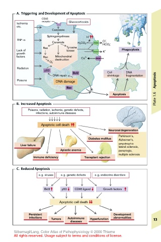
A.  Triggering  and Development of Apoptosis
                    CD95
        Ischemia    receptor    Glucocorticoids
        etc.
                        Caspases
                      Sphingomyelinase  H +
         TNF-α                                  –
                        Ceramide               Cl ,
                                    H 2 CO 3      –
                                               HCO 3
                    Ras        Tyrosine
        Lack of                kinase          K +     Phagocytosis
        growth
        factors     Rac   Mitochondrial  Ca 2+
                        – destruction
                       O 2
         Radiation
                     p53
                                  Bcl2           Cell    DNA
                         DNA repair              shrinkage  fragmentation
         Poisons          DNA damage                                   Apoptosis
                               Bax
                                                   Apoptosis
        B.  Increased Apoptosis                                        Plate 1.6
              Poisons, radiation, ischemia, genetic defects,
                 infections, autoimmune diseases

                   Apoptotic cell death
                                                  Neuronal degeneration
                                                      Parkinson’s,
                                         Diabetes mellitus  Alzheimer’s,
          Liver failure                               amyotrophic
                                                      lateral sclerosis,
                            Aplastic anemia           paraplegia,
                                                      multiple sclerosis
                Immune deficiency      Transplant rejection
        C.  Reduced Apoptosis
                  e.g. viruses  e.g. genetic defects  e.g. endocrine disorders


                     Bcl2    p53  CD95 ligand  Growth factors


                            Apoptotic cell death

              Persistent                            Development
              infections  Tumors  Autoimmune  Hyperfunction  abnormalities  13
                                diseases

       Silbernagl/Lang, Color Atlas of Pathophysiology © 2000 Thieme
       All rights reserved. Usage subject to terms and conditions of license.