
Page 296 - Color Atlas Of Pathophysiology (S Silbernagl Et Al, Thieme 2000)
P. 296
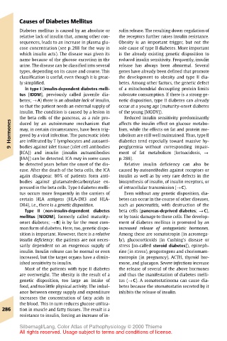
Causes of Diabetes Mellitus
       Diabetes mellitus is caused by an absolute or  sulin release. The resulting down-regulation of
       relative lack of insulin that, among other con-  the receptors further raises insulin resistance.
       sequences, leads to an increase in plasma glu-  Obesity is an important trigger, but not the
       cose concentration (see p. 288 for the way in  sole cause of type II diabetes. More important
       which insulin acts). The disease was given its  is the already existing genetic disposition to
       name because of the glucose excretion in the  reduced insulin sensitivity. Frequently, insulin
       urine. The disease can be classified into several  release has always been abnormal. Several
       types, depending on its cause and course. This  genes have already been defined that promote
       classification is useful, even though it is great-  the development to obesity and type II dia-
       ly simplified.                  betes. Among other factors, the genetic defect
         In type I (insulin-dependent diabetes melli-  of a mitochondrial decoupling protein limits
       tus [IDDM], previously called juvenile dia-  substrate consumption. If there is a strong ge-
       betes; → A) there is an absolute lack of insulin,  netic disposition, type II diabetes can already
       so that the patient needs an external supply of  occur at a young age (maturity-onset diabetes
       insulin. The condition is caused by a lesion in  of the young [MODY]).
       the beta cells of the pancreas, as a rule pro-  affects the insulin effect on glucose metabo-
                                        Reduced insulin sensitivity predominantly
    Hormones  may, in certain circumstances, have been trig-  lism, while the effects on fat and protein me-
       duced by an autoimmune mechanism that
                                       tabolism are still well maintained. Thus, type II
       gered by a viral infection. The pancreatic islets
                                       perglycemia without corresponding impair-
       bodies against islet tissue (islet cell antibodies
    9  are infiltrated by T lymphocytes and autoanti-  diabetics tend especially toward massive hy-
       [ICA]) and insulin (insulin autoantibodies  ment of fat metabolism (ketoacidosis, →
       [IAA]) can be detected. ICA may in some cases  p. 288).
       be detected years before the onset of the dis-  Relative insulin deficiency can also be
       ease. After the death of the beta cells, the ICA  caused by autoantibodies against receptors or
       again disappear. 80% of patients form anti-  insulin as well as by very rare defects in the
       bodies against glutamatedecarboxylase ex-  biosynthesis of insulin, of insulin receptors, or
       pressed in the beta cells. Type I diabetes melli-  of intracellular transmission (→ C).
       tus occurs more frequently in the carriers of  Even without any genetic disposition, dia-
       certain HLA antigens (HLA-DR3 and HLA-  betes can occur in the course of other diseases,
       DR4), i.e., there is a genetic disposition.  such as pancreatitis, with destruction of the
         Type II (non-insulin-dependent diabetes  beta cells (pancreas-deprived diabetes; → C),
       mellitus [NIDDM], formerly called maturity-  or by toxic damage to these cells. The develop-
       onset diabetes; → B) is by far the most com-  ment of diabetes mellitus is promoted by an
       mon form of diabetes. Here, too, genetic dispo-  increased release of antagonistic hormones.
       sition is important. However, there is a relative  Among these are somatotropin (in acromega-
       insulin deficiency: the patients are not neces-  ly), glucocorticoids (in Cushing’s disease or
       sarily dependent on an exogenous supply of  stress [so-called steroid diabetes]), epineph-
       insulin. Insulin release can be normal or even  rine (in stress), progestogens and choriomam-
       increased, but the target organs have a dimin-  motropin (in pregnancy), ACTH, thyroid hor-
       ished sensitivity to insulin.   mone, and glucagon. Severe infections increase
         Most of the patients with type II diabetes  the release of several of the above hormones
       are overweight. The obesity is the result of a  and thus the manifestation of diabetes melli-
       genetic disposition, too large an intake of  tus (→ C). A somatostatinoma can cause dia-
       food, and too little physical activity. The imbal-  betes because the stomatostatin secreted by it
       ance between energy supply and expenditure  inhibits the release of insulin.
       increases the concentration of fatty acids in
       the blood. This in turn reduces glucose utiliza-
  286  tion in muscle and fatty tissues. The result is a
       resistance to insulin, forcing an increase of in-
       Silbernagl/Lang, Color Atlas of Pathophysiology © 2000 Thieme
       All rights reserved. Usage subject to terms and conditions of license.