
Page 253 - ACCCN's Critical Care Nursing
P. 253
230 P R I N C I P L E S A N D P R A C T I C E O F C R I T I C A L C A R E
The final compensatory mechanism to be activated is the exists when the ventricle has an ejection fraction of less
neurohormonal response which takes days to be activated. than 40%, resulting in increased end-diastolic volume
64
This response involves the activation of vasopressin and and increased intraventricular pressure. The left atrium is
atrial natriuretic peptide (ANP). Vasopressin is a potent unable to empty into the left ventricle adequately and
vasoconstrictor and also an antidiuretic hormone. ANP is pressure in the left atrium rises. This pressure is reflected in
important in the regulation of cardiovascular volume the pulmonary veins and causes pulmonary congestion.
homeostasis. It is released from the atria in response to When pulmonary venous congestion exceeds 20 mmHg,
atrial stretching due to an increased circulating blood fluid moves into the pulmonary interstitium. Raised pul-
volume. ANP blocks the effect of the sympathetic nervous monary interstitial pressure reduces pulmonary compli-
system, RAAS and vasopressin. It reduces tachycardia via ance, increases the work of breathing and is experienced
the baroreceptors and reduces circulating blood volume by the patient as shortness of breath. Increased blood
by increasing salt and water excretion in the kidneys. volume in the lung also initiates shallow, rapid breathing
Plasma ANP is increased in acute heart failure but depleted and the sensation of breathlessness. Patients also experi-
in chronic heart failure. ence orthopnoea (dyspnoea while lying flat) and paroxys-
Whilst in the healthy heart, these compensatory mecha- mal nocturnal dyspnoea (PND), because when lying,
nisms would result in an adequate cardiac output, in blood is redistributed from gravity-dependent areas of the
heart failure they do not, depending on the aetiology. In body to the lung. Sitting upright or standing, and sleeping
64
ischaemic heart failure the damaged myocardium is with additional pillows, relieves breathlessness at night.
unable to respond adequately to the Frank-Starling Acute pulmonary oedema results when pulmonary capil-
response and ventricular remodelling develops. Heart lary pressure exceeds approximately 30 mmHg, and then
failure caused by hypertension or valvular heart disease fluid from the vessels begins to leak into the alveoli (see
results in persistent pressure or volume overload which Figure 10.10). This fluid leak decreases the area available
63
is exacerbated by the Frank-Starling response and sympa- for normal gas exchange and severe shortness of breath
thetic nervous system compensatory mechanisms. This results, often accompanied by pink, frothy sputum and
causes ventricular remodelling and depletion of norepi- noisy respirations. This causes patients to experience severe
nephrine and a reduction of inotropic response to the anxiety and decreased oxygen levels. Pulmonary oedema
cardiac sympathetic nervous system. These all exacerbate is a medical emergency and requires urgent treatment.
the reduction in circulating blood volume and kidney In addition to pulmonary symptoms, patients with left
perfusion. Many patients with heart failure often have a ventricular failure experience signs and symptoms related
high plasma renin activity due to the continual activation to decreased left ventricular output, including weakness,
of the RAAS compensatory mechanism.
fatigue, difficulty in concentrating and decreased exercise
In heart failure patients the inadequate cardiac output tolerance. These symptoms may be present for some time
results in signs and symptoms of hypoperfusion (oliguria, before an accurate diagnosis of heart failure is made,
cognitive impairment and cold peripheries) and conges- because they are non-specific and are consistent with
tion of the venous and pulmonary systems (acute pulmo- other diagnoses such as depression. Other signs that are
nary oedema, dyspnoea, hypoxaemia, peripheral oedema useful in diagnosis include the presence of S3 (ventricular
and liver congestion). Classification of signs and symp- gallop), crackles over lung fields that do not clear with a
toms is usually considered in the context of left or right cough, cardiomegaly and the presence of pulmonary
ventricular failure. vessels on chest X-ray.
LEFT VENTRICULAR FAILURE RIGHT VENTRICULAR FAILURE
Left ventricular failure (LVF), compared with other forms Right ventricular failure (RVF) does not usually occur in
of heart failure, is characterised by breathlessness, orthop- isolation, except in the presence of severe lung disease,
noea and paroxysmal nocturnal dyspnoea, irritating such as chronic obstructive pulmonary disease, pulmo-
60
cough and fatigue (see Table 10.4). Left ventricular failure nary hypertension or a massive pulmonary embolus. In
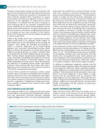
TABLE 10.4 Clinical manifestations of failure of right and left sides of the heart
Left ventricular failure Right ventricular failure
Signs Symptoms Signs Symptoms
Tachypnoea Dyspnoea Peripheral oedema Fatigue
Tachycardia Orthopnoea Raised jugular venous pressure Weight gain
Bibasal crackles Paroxysmal nocturnal dyspnoea Raised central venous pressure Anorexia
Haemoptysis Fatigue Ascites
Cough Nocturia Hepatomegaly
Pulmonary oedema
Raised pulmonary artery pressure
S3 heart sound