
Page 254 - ACCCN's Critical Care Nursing
P. 254
Cardiovascular Alterations and Management 231
Alveolus
Proteins
A Capillary Lymphatics B
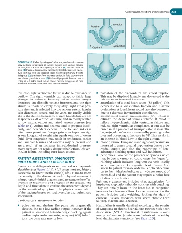
FIGURE 10.10 Pathophysiology of pulmonary oedema. As pulmo-
nary oedema progresses, it inhibits oxygen and carbon dioxide
exchange at the alveolar–capillary interface. (A) Normal relation-
ship. (B) Increased pulmonary capillary hydrostatic pressure causes
fluid to move from the vascular space into the pulmonary intersti-
tial space. (C) Lymphatic flow increases and pulls fluid back into the
vascular or lymphatic space. (D) Failure of lymphatic flow and wors-
ening of left-sided heart failure causes further movement of fluid C D
106
into the interstitial space and then into the alveoli.
this case, right ventricular failure is due to resistance to ● palpation of the praecordium and apical impulse:
outflow. The right ventricle can adapt to fairly large This may be displaced laterally and downward to the
changes in volume; however, when cardiac output left due to an increased heart size.
decreases, end-diastolic volume increases, and the right ● auscultation of a third heart sound (S3 gallop): This
atrium is unable to empty adequately. Right atrial pres- occurs due to a low ejection fraction and diastolic
sure rises and is reflected into the venous system. Jugular dysfunction. A fourth heart sound may also be present
vein distension occurs, and the veins are usually visible due to a decrease in ventricular compliance.
above the clavicle. Symptoms of right heart failure are not ● assessment of jugular venous pressure (JVP): This is to
as specific as left ventricular failure, and are mostly related estimate the degree of venous volume. If raised it
to low cardiac output and raised venous pressure (see reflects hypervolaemia, right ventricular failure, and
Table 10.4). Ascites and oedema tend to progress insidi- reduced right ventricular compliance. It can also be
ously, and dependent oedema in the feet and ankles is raised in the presence of tricuspid valve disease. The
often most prominent. Weight gain is an important sign hepatojugular reflex is also assessed by pressing on the
as one kilogram of weight gain equals one litre of excess liver and observing an increase in JVP. This results in
fluid. Liver congestion may result in tenderness, ascites an increase in blood flow to the right atrium.
and jaundice. Nausea and anorexia may be present and ● blood pressure: Lying and standing blood pressure are
are a result of an increased intra-abdominal pressure. measured to assess postural hypotension due to a low
Many signs are not readily distinguishable from left ven- cardiac output and also the prescribing of beta-
tricular failure, including extra heart sounds. adrenergic blocking agents and ACE inhibitors.
● peripheries: Look for the presence of cyanosis which
PATIENT ASSESSMENT, DIAGNOSTIC may be due to vasoconstriction. Assess the fingers for
PROCEDURES AND CLASSIFICATION clubbing which indicates long-term cyanosis usually
Assessment and diagnosis are summarised in a diagnostic as a consequence of congenital heart disease. Also
algorithm (see Figure 10.11). A full assessment and history assess the patient for ankle oedema. Peripheral oedema
is essential to determine the cause(s) of CHF and to assess up to the midcalves indicates a moderate amount of
the severity of the disease. A careful physical assessment excess fluid and the patient may require a bolus dose
is important for initial diagnosis and to evaluate the effec- of diuretic medication.
tiveness of treatments and progress of the disease. The Pulmonary assessment includes chest auscultation for
depth and time taken to conduct the assessment depend inspiratory crepitations that do not clear with coughing.
on the severity of symptoms. The physical examination They are initially heard in the bases but as congestion
of the patient focuses on cardiovascular and pulmonary increases they become diffuse. General assessment of the
assessment. patient includes daily weighing, looking for signs of
cachexia (usually associated with severe chronic heart
Cardiovascular assessment includes: failure), anaemia and dizziness.
● pulse rate and rhythm: The pulse rate is generally Heart failure is usually classified according to the severity
elevated due to a low cardiac output. However, if the of symptoms. In chronic heart failure, the New York Heart
patient is prescribed beta-adrenergic blocking agents Association (NYHA) Functional Classification is com-
and/or angiotensin converting enzyme (ACE) inhibi- monly used to classify patients on the basis of the activity
tors, the pulse rate may be low. level that initiates symptoms (see Table 10.5).