
Page 369 - ACCCN's Critical Care Nursing
P. 369
346 P R I N C I P L E S A N D P R A C T I C E O F C R I T I C A L C A R E
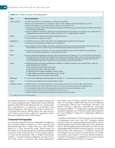
TABLE 13.7 Guide to normal CXR interpretation 65
Item Recommendation
Technical issues ● Check X-ray belongs to correct patient; note date and time of film.
● Ensure you are viewing the X-ray correctly (i.e. right and left markings correspond to thoracic structures).
● Determine whether X-ray was taken supine or erect, and whether PA or AP.
● Check X-ray was taken at full inspiration (posterior aspects of 9th/10th ribs and anterior aspects of 5th/6th ribs should
be visible above diaphragm).
● Note the penetration of the film: dark films are overpenetrated and may require a strong light to view; white films are
underpenetrated; good penetration will allow visualisation of the vertebrae behind the heart.
Bones ● Check along each rib from vertebral origin, looking for fractures.
● Ensure clavicles and scapulas are intact.
Mediastinum ● Check for presence of trachea and identify carina (approximately level of 5th–6th vertebrae).
● Check width of mediastinum: should not be more than 8 cm.
Apex ● Ensure blood vessels are visible in both apices, particularly looking to rule out pneumothoraces that present as clear
black shading on the X-ray. Erect X-rays are essential to facilitate visibility of pneumothoraces.
Hilum ● Check for prominence of vessels in this region: it generally indicates vascular abnormalities such as pulmonary
oedema or pulmonary hypertension, or congestive heart failure.
Heart ● Cardiac silhouette should be not more than 50% of the diameter of the thorax, with 3 of heart shadow to the right of
1
the vertebrae and 3 of shadow to the left of the vertebrae; this positioning helps to rule out a tension pneumothorax.
2
It should be noted that, post-cardiac surgery, if the mediastinum is left open the heart may appear wider than this;
also in AP films this may be the case due to the plate being further away from the heart.
Lung ● Identify the lobes of the lungs and determine if infiltrate or collapse is present in one or more of them. Lobes are
approximately located as follows:
● left upper lobe occupies upper half of lung;
● left lower lobe occupies lower half of lung;
● right lower lobe occupies costophrenic portion of lung;
● right middle lobe occupies cardiophrenic portion of lung;
● right upper lobe occupies upper portion of lung.
Diaphragm ● Check levels of diaphragm: right diaphragm will normally be 1–2 cm above the left diaphragm to accommodate the liver.
Gastric ● Check for pneumoperitoneum and dilated loops of bowel.
Catheters and lines ● Identify distal end of endotracheal tube and ensure above the carina (i.e. not in the right main bronchus).
● Trace nasogastric tube along length and ensure tip is in stomach, or below stomach if nasoenteric tube.
● Check position of intra-aortic balloon pump and ensure it is in the descending thoracic aorta.
● Trace all central catheters and ensure distal tip in correct location.
● Identify other lines (e.g. intercostal catheters, pacing wires) and note location.
PA = posterior-anterior; AP = anterior-posterior.
no need for transport of a critically ill patient outside the renal failure may preclude a patient from receiving con-
ICU, and it is radiation-free. Ultrasound is most useful for trast. CT scanning is useful in the detection and diagnosis
patients with fluid in the pleural space (i.e. pleural effu- of pulmonary, pleural and mediastinal disorders (e.g
sion, haemothorax or empyema), as it provides more pleural effusion, empyema, haemothorax, atelectasis,
73
70
detailed diagnostic information than chest X-rays alone; pneumonia, ARDS). CT pulmonary angiography (CTPA)
it estimates the volume of fluid present, the exact location produces a detailed view of blood vessels and is therefore
of the fluid, and provides a guide for aspirating a fluid- the most definitive method for diagnosing pulmonary
filled area or the placement of chest tubes. 71 embolism. 74
A significant limitation of CT scanning is that the patient
Computed Tomography is transported away from the ICU. Transport usually
Computed tomography (CT) is a diagnostic investigation requires at least two appropriately trained staff to accom-
that provides greater specificity in chest anatomy and pany the patient and involves added risk to the critically ill
pathophysiology than a plain CXR, as it uses multiple patient. Detailed planning by the health care team (includ-
beams in a circle around the body. These beams are ing imaging staff) includes ventilator support, monitoring
directed to a specific area of the body and provide detailed, requirements and maintenance of infusions during the
consecutive cross-sectional slices of the scanned regions. scanning period. See Chapter 6 for discussion of in-hospi-
CT scans can be performed with or without intravenous tal transfers, and Chapter 22 for inter-hospital transport.
72
contrast. Contrast improves diagnostic precision but is Portable CT scanners are available in some centres, but the
used with caution in patients with renal impairment; image quality is inferior to fixed CT scanners. 75