
Page 368 - ACCCN's Critical Care Nursing
P. 368
Respiratory Assessment and Monitoring 345
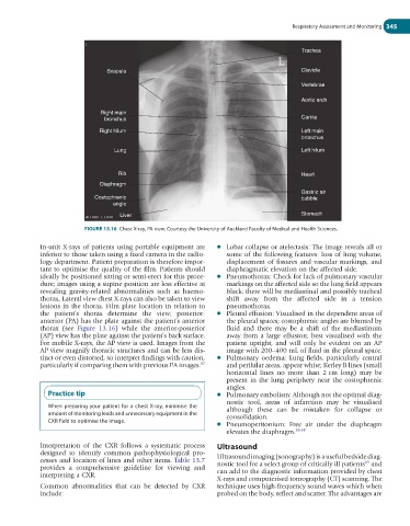
Trachea
Scapula Clavicle
Vertebrae
Aortic arch
Right main
bronchus Carina
Right hilum Left main
bronchus
Lung Left hilum
Rib Heart
Diaphragm
Gastric air
Costophrenic bubble
angle
Liver Stomach
FIGURE 13.16 Chest X-ray, PA view. Courtesy the University of Auckland Faculty of Medical and Health Sciences.
In-unit X-rays of patients using portable equipment are ● Lobar collapse or atelectasis: The image reveals all or
inferior to those taken using a fixed camera in the radio- some of the following features: loss of lung volume,
logy department. Patient preparation is therefore impor- displacement of fissures and vascular markings, and
tant to optimise the quality of the film. Patients should diaphragmatic elevation on the affected side.
ideally be positioned sitting or semi-erect for this proce- ● Pneumothorax: Check for lack of pulmonary vascular
dure; images using a supine position are less effective at markings on the affected side so the lung field appears
revealing gravity-related abnormalities such as haemo- black; there will be mediastinal and possibly tracheal
thorax. Lateral view chest X-rays can also be taken to view shift away from the affected side in a tension
lesions in the thorax. Film plate location in relation to pneumothorax.
the patient’s thorax determine the view; posterior- ● Pleural effusion: Visualised in the dependent areas of
anterior (PA) has the plate against the patient’s anterior the pleural spaces; costophrenic angles are blunted by
thorax (see Figure 13.16) while the anterior-posterior fluid and there may be a shift of the mediastinum
(AP) view has the plate against the patient’s back surface. away from a large effusion; best visualised with the
For mobile X-rays, the AP view is used. Images from the patient upright, and will only be evident on an AP
AP view magnify thoracic structures and can be less dis- image with 200–400 mL of fluid in the pleural space.
tinct or even distorted, so interpret findings with caution, ● Pulmonary oedema: Lung fields, particularly central
particularly if comparing them with previous PA images. 67 and perihilar areas, appear white; Kerley B lines (small
horizontal lines no more than 2 cm long) may be
present in the lung periphery near the costophrenic
angles.
Practice tip ● Pulmonary embolism: Although not the optimal diag-
nostic tool, areas of infarction may be visualised
When preparing your patient for a chest X-ray, minimise the although these can be mistaken for collapse or
amount of monitoring leads and unnecessary equipment in the consolidation.
CXR field to optimise the image.
● Pneumoperitonium: Free air under the diaphragm
elevates the diaphragm. 66,68
Interpretation of the CXR follows a systematic process Ultrasound
designed to identify common pathophysiological pro-
cesses and location of lines and other items. Table 13.7 Ultrasound imaging (sonography) is a useful bedside diag-
69
provides a comprehensive guideline for viewing and nostic tool for a select group of critically ill patients and
interpreting a CXR. can add to the diagnostic information provided by chest
X-rays and computerised tomography (CT) scanning. The
Common abnormalities that can be detected by CXR technique uses high-frequency sound waves which when
include: probed on the body, reflect and scatter. The advantages are