
Page 364 - ACCCN's Critical Care Nursing
P. 364
Respiratory Assessment and Monitoring 341
Capnography is recommended as a standard component
of respiratory monitoring in intubated and mechanically
41
ventilated patients in the ICU, during transport of a
D
40 critically ill patient and during anaesthesia. 43
42
C
PCO 2 (mm Hg) 20 B VENTILATION MONITORING
Mechanical ventilation is a common intervention in ICU
for patients with respiratory failure or who require respi-
ratory support. Advances in ventilation technology have
led to an increased ability to monitor many ventilator
parameters. A detailed understanding of mechanical ven-
tilation principles and functions enables patient data to
A be interpreted accurately and managed appropriately.
0 Chapter 15 provides a detailed discussion of mechanical
ventilation, including ventilation monitoring, airway
pressures (peak airway pressure, plateau pressure and
Expiration Inspiration positive end-expiratory pressure) and waveforms and
Time loop displays.
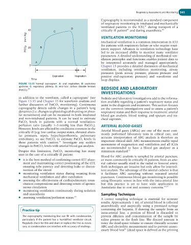
FIGURE 13.15 Normal capnogram. A: end inspiration; B: expiratory
upstroke; C: expiratory plateau; D: end-tidal carbon dioxide tension BEDSIDE AND LABORATORY
39
(PetCO 2 ).
INVESTIGATIONS
1
in addition to the waveform, called a capnogram (see Bedside and laboratory investigations add to the informa-
Figure 13.15 and Chapter 15 for waveform analysis and tion available regarding a patient’s respiratory status and
further discussion of PetCO 2 monitoring). Continuous assist in the diagnosis and treatment. This section focuses
capnography detects subtle changes in a patient’s lung on the common investigations used to assess a patient’s
dynamics (i.e. changes to physiological shunting or alveo- respiratory status and their response to treatment: arterial
lar recruitment) and can be measured in both intubated blood gas analysis; blood testing; and sputum and tra-
and non-intubated patients. It can be used to estimate cheal aspirates.
PaCO 2 levels in patients with a normal ventilation-
perfusion ratio (usually 1–5 mmHg less than PaCO 2 ). ARTERIAL BLOOD GASES
However, levels are affected by conditions common in the
critically ill (e.g. low cardiac output states, elevated alveo- Arterial blood gases (ABGs) are one of the most com-
lar pressures, sepsis, hypo/hyperthermia, pulmonary monly performed laboratory tests in critical care, and
embolism), so use PetCO 2 to estimate PaCO 2 levels in accurate interpretation of ABG analysis is therefore an
38
these patients with caution. Investigate any sudden important clinical skill. ABG measurements enable rapid
changes in PetCO 2 levels with arterial blood gas analysis. assessment of oxygenation and ventilation and all ICUs
are recommended to have a blood gas analyser as a
Despite this limitation, PetCO 2 monitoring has many minimum standard. 41
uses in the care of a critically ill patient:
Blood for ABG analysis is sampled by arterial puncture,
● it is the best method of confirming correct ETT place- or more commonly in critically ill patients, from an arte-
ment and maintaining correct positioning of the ETT, rial catheter usually sited in the radial or femoral artery.
ensuring tube patency and detecting leaks or discon- Both techniques are invasive but only allow for intermit-
nection of the circuit tent analysis. The advantage of the arterial catheter is that
● monitoring ventilation status during weaning from it facilitates ABG sampling without repeated arterial
mechanical ventilation and after extubation punctures. Continuous blood gas monitoring is possible
● assessing the effectiveness of cardiopulmonary resus- using fibreoptic sensor in-line with the intra-arterial line
citation compressions and detecting return of sponta- but this practice is yet to have wide application in
neous circulation Australasia due to cost and accuracy concerns. 44,45
● monitoring ventilation continuously during sedation
and anaesthesia Sampling Technique
● assessing ventilation/perfusion status. 40
A correct sampling technique is essential for accurate
results. Approximately 1 mL of arterial blood is collected
anaerobically and aseptically using a premixed syringe
Practice tip containing dry heparin. If drawing the sample from an
intra-arterial line, a portion of blood is discarded to
The capnography monitoring line can fill with condensation, prevent dilution and contamination of the sample by
particularly if the patient has a humidified ventilator circuit. saline present in the flush line. The discard amount is
Regularly check for this and drain or replace the line as neces- twice the dead space volume to ensure clinically accurate
sary, as condensation can interfere with accuracy of readings. ABG and electrolyte measurement and to prevent unnec-
essary blood loss (dead space is defined as the priming
46