
Page 365 - ACCCN's Critical Care Nursing
P. 365
342 P R I N C I P L E S A N D P R A C T I C E O F C R I T I C A L C A R E
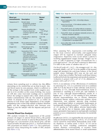
TABLE 13.4 Arterial blood gas normal values TABLE 13.5 Steps for arterial blood gas interpretation
Blood gas Normal Step Interpretation
measurements Description value 1 Assess oxygenation. PaO 2 < 60 mmHg indicates
Temperature (T) Patient’s body 37°C hypoxaemia.
temperature. Analyser 2 Assess the pH level. <7.36 indicates acidosis, >7.44
defaults to 37°C if not indicates alkalosis.
entered.
3 Assess PaCO 2 level. <35 mmHg indicates respiratory
Haemoglobin Samples should be fully Females:
(Hb) mixed so should be 115–165 g/L acidosis; >45 mmHg indicates respiratory alkalosis.
constantly agitated until Males: 4 Assess HCO 3 level. <22 indicates metabolic acidosis; >32
−
analysed. 130–180 g/L indicates metabolic alkalosis.
Acid–Base status Overall acidity or alkalinity 7.36–7.44 5 Assess pH, CO 2 and HCO 3 . Is there an acid–base
−
(pH) of blood. (36– disturbance and is it fully compensated, partially
44 mmol/L) compensated or uncompensated?
Carbon dioxide Partial pressure of arterial 35–45 mmHg 6 Assess other ABG results. Are they within normal limits
(CO 2 ) CO 2 . A potential acid. (4.7–6 kPa) for the patient?
Oxygen (O 2 ) Partial pressure of O 2 . 80–100 mmHg
Varies with age. (10.7–13.3 kPa)
−
Bicarbonate Standardised HCO 3 (actual 22–32 mmol/L When assessing PaO 2 , hypoxaemia (<60 mmHg) will
−
−
(HCO 3 ) HCO 3 minus the be the most common abnormality, and supplemental
−
HCO 3 produced by oxygen will be required to maintain adequate tissue
respiratory dysfunction) oxygenation. Hyperoxia rarely occurs unless a patient is
estimates true metabolic
function. An alkali or receiving supplemental oxygen therapy. Oxygen can be
base. toxic to cells if delivered at high concentrations for a
48
prolonged period. The pH level is assessed to determine
Base Excess (BE) Measures acid–base −3 to
balance. The number of +3 mmol/L if it falls on the acidic or alkaline side of 7.4.
molecules of acid or On the pH scale of 1–14 (1 = the strongest acid, 14 = the
base required to return 1
litre of blood to the strongest alkali), a pH of 7.4 is the middle of the normal
normal pH (7.4). range. pH measures the acid–base balance of the blood
+
sample, where Hydrogen (H ) ions are the acid and
Oxygen Haemoglobin saturation by 94.5–98.2% −
saturation oxygen in arterial blood HCO 3 is the base or buffer. The body’s acid–base balance
48
(SaO 2 ) is affected by both the respiratory and metabolic systems.
Acidaemia is present with a pH of <7.36; alkalaemia is
present with a pH of >7.44.
volume from sampling port to catheter tip; this differs PaCO 2 is an indicator of the effectiveness of ventilation
depending on the arterial line set up that is used). Arte- in removing CO 2 . CO 2 is a potential acid as it combines
rial blood exerts its own pressure, which is sufficient to with H 2 O in the blood to form carbonic acid (H 2 CO 3 ).
fill the syringe to the required level; active negative pres- Retention of CO 2 (through hypoventilation) leads to
+
sure is to be avoided, as this causes frothing. Any excess increased H resulting in a lower pH, and similarly a
air will also cause inaccurate readings and is expelled loss of CO 2 (through hyperventilation) results in a
49
before the syringe is capped with a hub, which prevents higher pH. A PaCO 2 of >45 mmHg (6 kPa) indicates
further contamination with air. The sample is analysed alveolar hypoventilation, due to chronic obstructive
within 10 minutes if not packed in ice, or within 60 pulmonary disease, asthma, pulmonary oedema, airway
minutes if iced; delays cause degradation of the sample. obstruction, over sedation, narcosis, drug overdose,
Degradation also occurs if the sample is shaken; therefore pain, neurological deficit or permissive hypercapnia in
50
gently roll the syringe/collection tube between your mechanically ventilated patients. Conversely, a PaCO 2
fingers to mix the sample with the heparin and prevent of <35 mmHg (4.7 kPa) reflects alveolar hyperventila-
clotting. 47 tion, and can be due to hypoxia, pain, anxiety, preg-
nancy, permissive hypocapnia in mechanically ventilated
Arterial Blood Gas Analysis patients or as a compensatory mechanism for metabolic
acidosis. 50
ABG analysis includes the measurement of the partial
−
pressure of oxygen in arterial blood (PaO 2 ), the partial Bicarbonate (HCO 3 ) is regulated by the renal system and
−
pressure of carbon dioxide in arterial blood (PaCO 2 ), the indicates metabolic functioning. A HCO 3 of < 22 mmol/L
hydrogen ion concentration of the blood (pH), and the can be caused by renal failure, ketoacidosis, lactic acido-
−
–
chemical buffer, bicarbonate (HCO 3 ). Normal values for sis, diarrhoea, or cardiac arrest. A HCO 3 of >32 can be
ABG parameters are listed in Table 13.4. Use a systematic caused by severe vomiting, continuous nasogastric
approach when interpreting the results of ABG analysis suction, diuretics, corticosteroids, or excessive citrate
(see Table 13.5). administration from stored blood or renal replacement