
Page 376 - ACCCN's Critical Care Nursing
P. 376
Respiratory Alterations and Management 353
● increased metabolic oxygen requirements may be
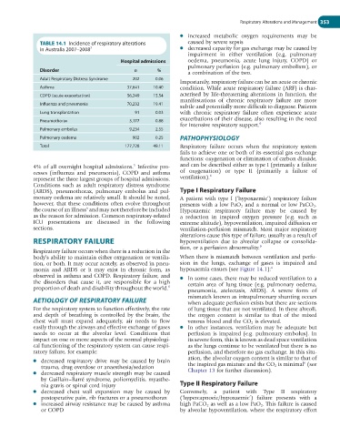
TABLE 14.1 Incidence of respiratory alterations caused by severe sepsis
in Australia 2007–2008 7 ● decreased capacity for gas exchange may be caused by
impairment in either ventilation (e.g. pulmonary
Hospital admissions oedema, pneumonia, acute lung injury, COPD) or
pulmonary perfusion (e.g. pulmonary embolism), or
Disorder n % a combination of the two.
Adult Respiratory Distress Syndrome 202 0.06
Importantly, respiratory failure can be an acute or chronic
Asthma 37,641 10.40 condition. While acute respiratory failure (ARF) is char-
COPD (acute exacerbation) 56,249 15.54 acterised by life-threatening alterations in function, the
manifestations of chronic respiratory failure are more
Influenza and pneumonia 70,232 19.41
subtle and potentially more difficult to diagnose. Patients
Lung transplantation 91 0.03 with chronic respiratory failure often experience acute
Pneumothorax 3,177 0.88 exacerbations of their disease, also resulting in the need
for intensive respiratory support. 6
Pulmonary embolus 9,234 2.55
Pulmonary oedema 902 0.25 PATHOPHYSIOLOGY
Total 177,728 49.11 Respiratory failure occurs when the respiratory system
fails to achieve one or both of its essential gas exchange
functions: oxygenation or elimination of carbon dioxide,
5
4% of all overnight hospital admissions. Infective pro- and can be described either as type I (primarily a failure
cesses (influenza and pneumonia), COPD and asthma of oxygenation) or type II (primarily a failure of
6
represent the three largest groups of hospital admissions. ventilation).
Conditions such as adult respiratory distress syndrome
(ARDS), pneumothorax, pulmonary embolus and pul- Type I Respiratory Failure
monary oedema are relatively small. It should be noted, A patient with type I (‘hypoxaemic’) respiratory failure
however, that these conditions often evolve throughout presents with a low PaO 2 and a normal or low PaCO 2 .
the course of an illness and may not therefore be included Hypoxaemic respiratory failure may be caused by
6
as the reason for admission. Common respiratory-related a reduction in inspired oxygen pressure (e.g. such as
ICU presentations are discussed in the following extreme altitude), hypoventilation, impaired diffusion or
sections. ventilation-perfusion mismatch. Most major respiratory
alterations cause this type of failure, usually as a result of
RESPIRATORY FAILURE hypoventilation due to alveolar collapse or consolida-
tion, or a perfusion abnormality. 6
Respiratory failure occurs when there is a reduction in the
body’s ability to maintain either oxygenation or ventila- When there is mismatch between ventilation and perfu-
tion, or both. It may occur acutely, as observed in pneu- sion in the lungs, exchange of gases is impaired and
monia and ARDS or it may exist in chronic form, as hypoxaemia ensues (see Figure 14.1): 6
observed in asthma and COPD. Respiratory failure, and ● In some cases, there may be reduced ventilation to a
the disorders that cause it, are responsible for a high certain area of lung tissue (e.g. pulmonary oedema,
proportion of death and disability throughout the world. 6
pneumonia, atelectasis, ARDS). A severe form of
AETIOLOGY OF RESPIRATORY FAILURE mismatch known as intrapulmonary shunting occurs
when adequate perfusion exists but there are sections
For the respiratory system to function effectively, the rate of lung tissue that are not ventilated. In these alveoli,
and depth of breathing is controlled by the brain, the the oxygen content is similar to that of the mixed
chest wall must expand adequately, air needs to flow venous blood and the CO 2 is elevated.
easily through the airways and effective exchange of gases ● In other instances, ventilation may be adequate but
needs to occur at the alveolar level. Conditions that perfusion is impaired (e.g. pulmonary embolus). In
impact on one or more aspects of the normal physiologi- its severe form, this is known as dead space ventilation
cal functioning of the respiratory system can cause respi- as the lungs continue to be ventilated but there is no
ratory failure, for example: perfusion, and therefore no gas exchange. In this situ-
ation, the alveolar oxygen content is similar to that of
● decreased respiratory drive may be caused by brain 6
trauma, drug overdose or anaesthesia/sedation the inspired gas mixture and the CO 2 is minimal (see
● decreased respiratory muscle strength may be caused Chapter 13 for further discussion).
by Guillain–Barré syndrome, poliomyelitis, myasthe-
nia gravis or spinal cord injury Type II Respiratory Failure
● decreased chest wall expansion may be caused by Conversely, a patient with Type II respiratory
postoperative pain, rib fractures or a pneumothorax (‘hypercapnoeic/hypoxaemic’) failure presents with a
● increased airway resistance may be caused by asthma high PaCO 2 as well as a low PaO 2 . This failure is caused
or COPD by alveolar hypoventilation, where the respiratory effort