
Page 378 - ACCCN's Critical Care Nursing
P. 378
Respiratory Alterations and Management 355
or tidal volume to maintain a specified PaCO 2 . One method should be considered for all ventilated patients.
concern that often arises, particularly with patients who The approach may result in tolerance of higher PaCO 2
require high concentrations of oxygen, is the risk of than normal in patients presenting with acute lung
oxygen toxicity. The link between prolonged periods of injury or ARDS (see Chapter 15 for further
oxygen concentrations approaching 100% and oxidant discussion).
injuries in airways and lung parenchyma has been estab- Development of ventilator-associated respiratory muscle
lished, although mostly from animal research. Although weakness has been reported as a significant issue when
it remains unclear how these data apply to human popu- the respiratory muscles are rendered inactive through
lations, most consensus groups have argued that FiO 2 adjustment of ventilator settings and administration of
values less than 0.4 are safe for prolonged periods of time pharmacotherapy. While it is not yet possible to provide
and that FiO 2 values of greater than 0.8 should be avoided precise recommendations for interventions to avoid this,
6
if possible (see Chapter 15 for further discussion of clinicians are advised to select ventilator settings that
oxygenation). provide for some respiratory muscle use. 11
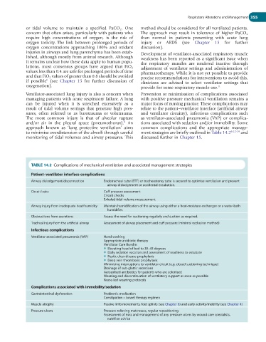
Ventilator-associated lung injury is also a concern when Prevention or minimisation of complications associated
managing patients with acute respiratory failure. A lung with positive pressure mechanical ventilation remains a
can be injured when it is stretched excessively as a major focus of nursing practice. These complications may
result of tidal volume settings that generate high pres- relate to the patient–ventilator interface (artificial airway
sures, often referred to as barotrauma or volutrauma. and ventilator circuitry), infectious complications such
The most common injury is that of alveolar rupture as ventilator-associated pneumonia (VAP) or complica-
6
and/or air in the pleural space (pneumothorax). An tions associated with sedation and/or immobility. Some
approach known as ‘lung protective ventilation’ aims common complications and the appropriate manage-
to minimise overdistension of the alveoli through careful ment strategies are briefly outlined in Table 14.2 6,12-14 and
monitoring of tidal volumes and airway pressures. This discussed further in Chapter 15.
TABLE 14.2 Complications of mechanical ventilation and associated management strategies
Patient–ventilator interface complications
Airway dislodgement/disconnection Endotracheal tube (ETT) or tracheostomy tube is secured to optimise ventilation and prevent
airway dislodgement or accidental extubation.
Circuit leaks Cuff pressure assessment
Circuit checks
Exhaled tidal volume measurement
Airway injury from inadequate heat/humidity Maintain humidification of the airway using either a heat-moisture exchanger or a water-bath
humidifier.
Obstructions from secretions Assess the need for suctioning regularly and suction as required.
Tracheal injury from the artificial airway Assessment of airway placement and cuff pressure (minimal occlusion method)
Infectious complications
Ventilator-associated pneumonia (VAP) Hand washing
Appropriate antibiotic therapy
Ventilator Care Bundle:
● Elevating head of bed to 30–45 degrees
● Daily sedation vacation and assessment of readiness to extubate
● Peptic ulcer disease prophylaxis
● Deep vein thrombosis prophylaxis
Minimising interruptions to ventilator circuit (e.g. closed suctioning technique)
Drainage of sub-glottic secretions
Aerosolised antibiotics for patients who are colonised
Weaning and discontinuation of ventilatory support as soon as possible
Nurse-led weaning protocols
Complications associated with immobility/sedation
Gastrointestinal dysfunction Prokinetic medication
Constipation – bowel therapy regimen
Muscle atrophy Passive limb movements, foot splints (see Chapter 6) and early activity/mobility (see Chapter 4)
Pressure ulcers Pressure-relieving mattresses, regular repositioning
Assessment of risks and management of any pressure ulcers by wound care specialists,
nutrition advice