
Page 377 - ACCCN's Critical Care Nursing
P. 377
354 P R I N C I P L E S A N D P R A C T I C E O F C R I T I C A L C A R E
Pure
shunt
V/Q = 0
52
Alveolar PCO 2 (mmHg) Decreasing V/Q Normal V/Q space
Pure
dead
Increasing V/Q
V/Q = ∞
0
45 150
Alveolar PO 2 (mmHg)
6
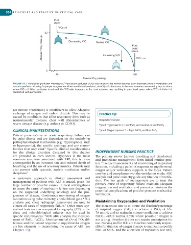
FIGURE 14.1 Ventilation-perfusion mismatches. Ventilation-perfusion (V/Q) ratio displays the normal balance (star) between alveolar ventilation and
vascular perfusion allowing for proper oxygenation. When ventilation is reduced, the V/Q ratio decreases, in the most extreme case resulting in pure shunt,
where V/Q = 0. When perfusion is reduced, the V/Q ratio increases, in the most extreme case resulting in pure dead space, where V/Q = infinite (∞).
(published with permission)
(or minute ventilation) is insufficient to allow adequate
exchange of oxygen and carbon dioxide. This may be Practice tip
caused by conditions that affect respiratory drive such as
neuromuscular diseases, chest wall abnormalities or Respiratory failure:
severe airways disease (e.g. asthma or COPD).
Type I (‘hypoxaemic’) = low PaO 2 and normal or low PaCO 2
CLINICAL MANIFESTATIONS Type II (‘hypercapnoeic’) = high PaCO 2 and low PaO 2
Patient presentations in acute respiratory failure can
be quite diverse and are dependent on the underlying
pathophysiological mechanism (e.g. hypercapnoea and/
or hypoxaemia), the specific aetiology and any comor-
bidities that may exist. Specific clinical manifestations
6
for the clinical disorders discussed in this chapter INDEPENDENT NURSING PRACTICE
are provided in each section. Dyspnoea is the most The primary survey (airway, breathing and circulation)
common symptom associated with ARF; this is often and immediate management form initial routine prac-
accompanied by an increased rate and reduced depth of tice. Frequent assessment and monitoring of respiratory
10
breathing and the use of accessory muscles. Patients may function, including a patient’s response to supplemental
also present with cyanosis, anxiety, confusion and/or oxygen and/or ventilatory support, is the focus. Patient
sleepiness. 4 comfort and compliance with the ventilation mode, ABG
A systematic approach to clinical assessment and analysis and pulse oximetry guide any titration of ventila-
management of patients with ARF is crucial, given the tion. The key goals of management are to treat the
large number of possible causes. Clinical investigations primary cause of respiratory failure, maintain adequate
to assess the cause of respiratory failure vary depending oxygenation and ventilation and prevent or minimise the
on the suspected underlying aetiology and the pro- potential complications of positive pressure mechanical
gression of disease. Continuous monitoring of oxygen ventilation.
saturation using pulse oximetry, arterial blood gas (ABG)
analysis and chest radiograph assessment are used in Maintaining Oxygenation and Ventilation
8
almost all cases of respiratory failure. Other more spe- The therapeutic aim is to titrate the fraction/percentage
cialised tests such as computed tomography (CT) of the of inspired oxygen (FiO 2 ) to achieve a PaO 2 of 65–
chest and microbiological cultures may be used in 70 mmHg and to maintain minute ventilation to achieve
6
9
specific circumstances. With ABG analysis, the measure- PaCO 2 within normal limits where possible. Oxygen is
ment of PaO 2 , PaCO 2 , Alveolar–arterial (A–a) PO 2 dif- not a drug, therefore it does not require prescription for
ference and the patient response to supplemental oxygen use. Nursing staff in ICU are therefore commonly respon-
are key elements in determining the cause of ARF (see sible for titration of oxygen therapy to maintain a specific
Chapter 13). PaO 2 or SpO 2 , and the alteration of respiratory rate and/