
Page 381 - ACCCN's Critical Care Nursing
P. 381
358 P R I N C I P L E S A N D P R A C T I C E O F C R I T I C A L C A R E
recommended systems that produce scores and assess
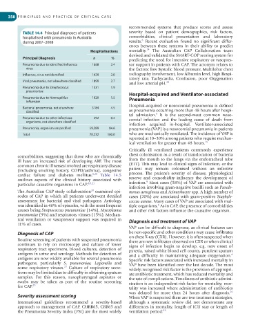
TABLE 14.4 Principal diagnoses of patients severity based on patient demographics, risk factors,
hospitalised with pneumonia in Australia comorbidities, clinical presentation and laboratory
6
during 2007–2008 results. Recent evaluation found no significant differ-
ences between these systems in their ability to predict
24
Hospitalisations mortality. The Australian CAP Collaboration team
devised and validated the SMART-COP scoring system for
Principal Diagnosis n % predicting the need for intensive respiratory or vasopres-
Pneumonia due to identified influenza 1668 2.4 sor support in patients with CAP. The acronym relates to
virus the factors: low Systolic blood pressure, Multilobar chest
Influenza, virus not identified 1429 2.0 radiography involvement, low Albumin level, high Respi-
ratory rate, Tachycardia, Confusion, poor Oxygenation
Viral pneumonia, not elsewhere classified 1899 2.7 25
and low arterial pH.
Pneumonia due to Streptococcus 1331 1.9
pneumoniae Hospital-acquired and Ventilator-associated
Pneumonia due to Haemophilus 1029 1.5 Pneumonia
influenzae
Hospital-acquired or nosocomial pneumonia is defined
Bacterial pneumonia, not elsewhere 3184 4.5
classified as pneumonia occurring more than 48 hours after hospi-
tal admission. It is the second-most common noso-
9
Pneumonia due to other infectious 292 0.4 comial infection and the leading cause of death from
organisms, not elsewhere classified
infection acquired in-hospital. Ventilator-associated
Pneumonia, organism unspecified 59,389 84.6 pneumonia (VAP) is a nosocomial pneumonia in patients
Total 70,232 100.0 who are mechanically ventilated. The incidence of VAP is
reported at 10–30% among patients who require mechan-
ical ventilation for greater than 48 hours. 26
Critically ill ventilated patients commonly experience
comorbidities, suggesting that those who are chronically chest colonisation as a result of translocation of bacteria
ill have an increased risk of developing ARF. The most from the mouth to the lungs via the endotracheal tube
common chronic illnesses involved are respiratory disease (ETT). This may lead to clinical signs of infection, or the
(including smoking history, COPD/asthma), congestive patient may remain colonised without an infective
cardiac failure and diabetes mellitus. 6,20 Table 14.5 process. The patient’s severity of disease, physiological
outlines aspects of the clinical history associated with reserve and comorbidity influence the development of
6
particular causative organisms in CAP. 6,9,21 infection. Most cases (58%) of VAP are associated with
infection involving gram-negative bacilli such as Pseudo-
20
The Australian CAP study collaboration examined epi- monas aeruginosa and Acinetobacter spp. A high number of
sodes of CAP in which all patients underwent detailed cases (20%) are associated with gram-positive Staphylo-
assessment for bacterial and viral pathogens. Aetiology coccus aureus. Many cases of VAP are associated with mul-
was identified in 46% of episodes, with the most frequent tiple organisms. As in CAP, the presence of comorbidities
6
causes being Streptococcus pneumoniae (14%), Mycoplasma and other risk factors influence the causative organism.
pneumoniae (9%) and respiratory viruses (15%). Mechan-
ical ventilation or vasopressor support was required in Diagnosis and treatment of VAP
11% of cases.
VAP can be difficult to diagnose, as clinical features can
Diagnosis of CAP be non-specific and other conditions may cause infiltrates
Routine screening of patients with suspected pneumonia on chest X-ray (CXR). However, it is often suspected when
there are new infiltrates observed on CXR or when clinical
continues to rely on microscopy and culture of lower signs of infection begin to develop, e.g. new onset of
respiratory tract specimens, blood cultures, detection of pyrexia, raised white blood cell counts, purulent sputum
antigens in urine and serology. Methods for detection of and a difficulty in maintaining adequate oxygenation.
6
antigens are now widely available for several pneumonia Specific risk factors associated with increased mortality in
pathogens, particularly S. pneumoniae, Legionella and VAP have been identified over the last decade. The most
22
some respiratory viruses. Culture of respiratory secre- widely-recognised risk factor is the provision of appropri-
tions may be limited due to difficulty in obtaining sputum ate antibiotic treatment, which has reduced mortality and
samples. For this reason, nasopharyngeal aspirates or the rate of complications. Timeliness of antibiotic admini-
swabs may be taken as part of the routine screening stration is an independent risk factor for mortality; mor-
for CAP. 23 tality was increased where administration of antibiotics
26
was delayed for more than 24 hours after diagnosis.
Severity assessment scoring When VAP is suspected there are two treatment strategies,
International guidelines recommend a severity-based although a systematic review did not demonstrate any
approach to management of CAP. CURB65, CRB65 and differences in mortality, length of ICU stay or length of
the Pneumonia Severity Index (PSI) are the most widely ventilation period: 19