
Page 395 - ACCCN's Critical Care Nursing
P. 395
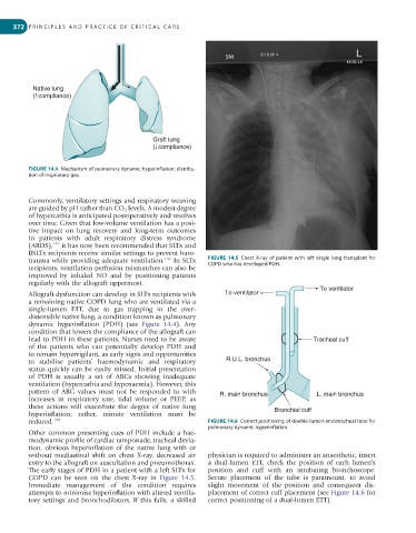
372  P R I N C I P L E S   A N D   P R A C T I C E   O F   C R I T I C A L   C A R E


           Native lung
           (  compliance)


                                              Graft lung
                                              (  compliance)


         FIGURE 14.4  Mechanism of pulmonary dynamic hyperinflation: distribu-
         tion of inspiratory gas.


         Commonly, ventilatory settings and respiratory weaning
         are guided by pH rather than CO 2  levels. A modest degree
         of hypercarbia is anticipated postoperatively and resolves
         over time. Given that low-volume ventilation has a posi-
         tive  impact  on  lung  recovery  and  long-term  outcomes
         in  patients  with  adult  respiratory  distress  syndrome
         (ARDS), 147  it has now been recommended that SLTx and
         BSLTx recipients receive similar settings to prevent baro-
         trauma while providing adequate ventilation. 142  In SLTx   FIGURE 14.5  Chest  X-ray  of  patient  with  left  single  lung  transplant  for
         recipients, ventilation perfusion mismatches can also be   COPD who has developed PDH.
         improved  by  inhaled  NO  and  by  positioning  patients
         regularly with the allograft uppermost.                                               To ventilator
         Allograft dysfunction can develop in SLTx recipients with   To ventilator
         a remaining native COPD lung who are ventilated via a
         single-lumen  ETT,  due  to  gas  trapping  in  the  over-
         distensible native lung, a condition known as pulmonary
         dynamic  hyperinflation  (PDH)  (see  Figure  14.4).  Any
         condition that lowers the compliance of the allograft can
         lead to PDH in these patients. Nurses need to be aware                              Tracheal cuff
         of  the  patients  who  can  potentially  develop  PDH  and
         to remain hypervigilant, as early signs and opportunities
         to  stabilise  patients’  haemodynamic  and  respiratory   R.U.L. bronchus
         status quickly can be easily missed. Initial presentation
         of  PDH  is  usually  a  set  of  ABGs  showing  inadequate
         ventilation (hypercarbia and hypoxaemia). However, this
         pattern  of  ABG  values  must  not  be  responded  to  with   R. main bronchus      L. main bronchus
         increases  in  respiratory  rate,  tidal  volume  or  PEEP,  as
         these  actions  will  exacerbate  the  degree  of  native  lung
         hyperinflation;  rather,  minute  ventilation  must  be                  Bronchial cuff
         reduced. 148                                         FIGURE 14.6  Correct positioning of double-lumen endotracheal tube for
                                                              pulmonary dynamic hyperinflation.
         Other common presenting cues of PDH include a hae-
         modynamic profile of cardiac tamponade, tracheal devia-
         tion, obvious hyperinflation of the native lung with or
         without  mediastinal  shift  on  chest  X-ray,  decreased  air   physician is required to administer an anaesthetic, insert
         entry to the allograft on auscultation and pneumothorax.   a  dual-lumen  ETT,  check  the  position  of  each  lumen’s
         The early stages of PDH in a patient with a left SLTx for   position  and  cuff  with  an  intubating  bronchoscope.
         COPD  can  be  seen  on  the  chest  X-ray  in  Figure  14.5.   Secure  placement  of  the  tube  is  paramount,  to  avoid
         Immediate  management  of  the  condition  requires   slight  movement  of  the  position  and  consequent  dis-
         attempts to minimise hyperinflation with altered ventila-  placement of correct cuff placement (see Figure 14.6 for
         tory settings and bronchodilators. If this fails, a skilled   correct positioning of a dual-lumen ETT).