
Page 394 - ACCCN's Critical Care Nursing
P. 394
Respiratory Alterations and Management 371
due to infection or hemidiaphragm paralysis secondary
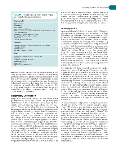
TABLE 14.14 Possible causes of low cardiac output in to phrenic nerve damage. Although BSLTx is usually per-
first week after lung transplantation formed without cardiopulmonary bypass, for those
patients who require cardiopulmonary bypass for surgery,
Cardiovascular it is recognised that there is a higher incidence of PGD
but management principles are essentially the same.
Hypovolaemia
Haemorrhage
Hypothermia
Acute myocardial infarction Nursing practice
Pulmonary venous or arterial anastomosis obstruction (embolism, Severity of allograft dysfunction is assessed by ABG analy-
clot, stitch, torsion) sis, respiratory function and patient comfort, chest X-ray,
Pulmonary embolism (thrombus or air)
Non-specific left ventricular dysfunction bronchoscopy and haemodynamic parameters. A careful
Arrhythmias balance in the management of haemodynamic, respira-
Coronary artery air embolism tory and renal status is vital in the first 12 hours, and their
Pulmonary optimisation should be achieved with inotropes (e.g.
adrenaline, noradrenaline) and limited and judicious use
Pulmonary dynamic inflation of native lung in single-lung of colloid fluids to ensure adequate end-organ perfusion
transplantation
Pneumothorax without causing pulmonary overload. Fluid management
Oversized pulmonary allograft should aim to keep filling pressures low to normal in
light of a recent retrospective review that found a high
Other
CVP (>7 mmHg) was associated with prolonged mechan-
141
Sepsis/infection (especially line or occult gut) ical ventilation and high mortality. Importantly, there
Sedatives was no evidence of renal complications associated with
Analgesics (especially epidural) 141
Transfusion reaction these low filling pressures. Fluid resuscitation should
Anaphylaxis include products to correct anaemia and preoperative low
Hyperacute rejection (rare) plasma protein levels. 142
For patients who have required intraoperative cardio-
pulmonary bypass, high doses of inotropes are often
Haemodynamic function can be compromised in the needed to overcome a transient relative hypovolaemia.
early postoperative phase due to cardiac and respiratory Additionally, gentle rewarming measures are needed to
problems; renal and gastrointestinal dysfunction is also re-establish normothermia in order to prevent haema-
prevalent. Long-term respiratory complications include tological and peripheral perfusion impairments associ-
airway anastomotic problems (stricture and dehiscence), ated with hypothermia. Slow rather than rapid rewarming,
suboptimal exercise performance, and chronic rejection and close monitoring of CI, CVP and PAWP should
manifesting as bronchiolitis obliterans syndrome. The minimise the development of pulmonary oedema at this
most important aspects of these complications are dis- time. For patients with allograft dysfunction accompa-
cussed below in relation to nursing practice, and Table nied by high pulmonary pressures, inhaled NO is useful
14.14 provides a summary. in decreasing high pulmonary pressures and intrapul-
monary shunting. 143,144 Ongoing nursing assessments of
Respiratory Dysfunction MAP, CI, PAP, PAWP, CVP and urine output guide and
Respiratory dysfunction within the first 24–48 hours evaluate haemodynamic therapeutic interventions (see
Chapter 9).
postoperatively is usually caused by primary graft
dysfunction (PGD), a syndrome characterised by non- To assess the causes and progress of allograft dysfunction,
specific alveolar damage, lung oedema and hypoxae- chest X-rays provide vital information about line place-
mia. 137 Primary graft dysfunction may be aggravated by ment, ETT position, lung expansion, lung size, position
factors associated with the donor (e.g. trauma, mechani- of the diaphragm and mediastinum and the presence of
cal ventilation, aspiration, pneumonia and hypotension), pneumothorax, oedema and atelectasis. 145 Allograft dys-
cold ischaemic storage, 137 inadequate preservation and function due to ischaemia-reperfusion injury appears on
disruption of pulmonary lymphatics. Clinical signs of chest X-rays as a rapidly-developing diffuse alveolar
PGD range from mild hypoxaemia with infiltrates on pattern of infiltration that is greater in the lower regions, 142
chest X-rays to severe ARDS requiring high-level ventila- most commonly seen on the first postoperative day but
138
tory support, pharmacological support and ECMO. may occur up to 72 hours following surgery. The presence
Australian researchers have shown a decrease in the sever- of rapidly worsening pulmonary infiltrates (especially if
ity and incidence of PGD following the implementation associated with low cardiac indices) should however
of an evidence-based guideline for managing patients’ prompt urgent echocardiography to assess cardiac func-
respiratory and haemodynamic status postoperatively. 139 tion and pulmonary venous anastamosis patency. Beyond
The guideline directs clinicians to minimise crystalloid 72 hours, alveolar and interstitial infiltration may indi-
fluids, use vasopressors as the first-line treatment to cate either acute rejection or an infective process. 146 This
maintain blood pressure if cardiac output is adequate information is combined with other respiratory and hae-
and use ARDSNet principles for ventilatory support. 139,140 modynamic data to inform appropriate collaborative
Respiratory dysfunction beyond 72 hours is likely to be interventions.