
Page 390 - ACCCN's Critical Care Nursing
P. 390
Respiratory Alterations and Management 367
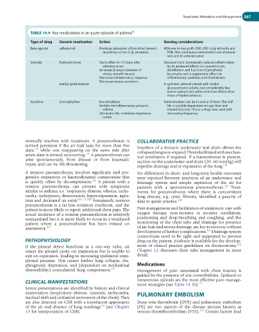
TABLE 14.9 Key medications in an acute episode of asthma 58
Type of drug Generic medication Action Nursing considerations
Beta-agonist salbutamol Produces relaxation of bronchial smooth MDI-one to two puffs (100–200 mcg) 4-hourly and
muscle by action at β 2 -receptors. PRN. Also continuous nebulisation via ultrasonic
neb and IV administration
Steroids hydrocortisone Starts effect 6–12 hours after Glucocorticoid dramatically reduces inflammation
administration. by its profound effects on concentration,
Increases β-responsiveness of distribution and function of peripheral
airway smooth muscle. leucocytes and a suppressive effect on
Decreases inflammatory response. inflammatory cytokines and chemokines.
Decreases mucus secretion.
methyl-prednisolone A synthetic adrenal steroid with similar
glucocorticoid activity, but considerably less
severe sodium and water retention effects than
those of hydrocortisone.
Xanthine aminophylline Bronchodilator Administration can be in oral or IV form. The half
Inhibits the inflammatory phase in life is variable dependent on age, liver and
asthma thyroid function. This is a drug now used with
Stimulates the medullary respiratory decreasing frequency
centre
normally resolves with treatment. A pneumothorax is COLLABORATIVE PRACTICE
termed persistent if the air leak lasts for more than five Insertion of a thoracic underwater seal drain allows the
days, while one reappearing on the same side after collapsed lung to re-expand. This is facilitated with mechan-
114
115
seven days is termed reoccurring. A pneumothorax can ical ventilation if required. If a haemothorax is present,
arise spontaneously, from disease or from traumatic suction on the underwater seal drain (20–60 mmHg) will
injury and can be life-threatening.
expedite drainage and re-expansion of the lung. 118
A tension pneumothorax involves significant and pro- No differences in short- and long-term health outcomes
gressive respiratory or haemodynamic compromise that were reported between insertion of an underwater seal
116
is quickly offset by decompression. A patient with a drainage system and simple aspiration of the air for
tension pneumothorax can present with symptoms patients with a spontaneous pneumothorax. Treat-
119
similar to asthma, i.e. ‘respiratory distress, wheeze, tachy- ments for pneumothorax where there is concomitant
cardia, tachypnoea, desaturation, hyper-expansion, agita- lung disease, e.g. cystic fibrosis, identified a paucity of
tion and decreased air entry.’ 117, p. 525 Fortunately, tension data to guide practice. 120
pneumothorax is a far less common condition, and the
patient is more likely to report additional chest pain. The Pain management and facilitation of respiratory care with
actual incidence of a tension pneumothorax is relatively oxygen therapy, non-invasive or invasive ventilation,
unexamined but it is more likely to occur in a ventilated positioning and deep-breathing and coughing, and the
patient where a pneumothorax has been missed on monitoring of the chest tube and drainage for presence
assessment. 117 of air-leak and serous drainage, are key to recovery without
121
development of further complications. Drainage system
connections need to be tight and supported to prevent
PATHOPHYSIOLOGY drag on the patient. Evidence is available for the develop-
121
If the pleural defect functions as a one-way valve, air ment of clinical practice guidelines on thoracostomy.
enters the pleural cavity on inspiration but is unable to Chapter 12 discusses chest tube management in more
exit on expiration, leading to increasing ipsilateral intra- detail.
pleural pressure. This causes further lung collapse, dia-
phragmatic depression, and (dependent on mediastinal Medications
distensibility) contralateral lung compression. 117 Management of pain associated with chest trauma is
guided by the presence of any comorbidities. Epidural or
CLINICAL MANIFESTATIONS intravenous opioids are the most effective pain manage-
121
ment strategies (see Table 14.10).
Severe presentations are identified by history and clinical
examination (respiratory distress, cyanosis, tachycardia, PULMONARY EMBOLISM
tracheal shift and unilateral movement of the chest). They
are also detected on CXR with a translucent appearance Deep vein thrombosis (DVT) and pulmonary embolism
118
of the air and absence of lung markings (see Chapter (PE) are two aspects of the disease process known as
13 for interpretation of CXR). venous thromboembolism (VTE). 122 Certain factors lead