
Page 459 - ACCCN's Critical Care Nursing
P. 459
436 P R I N C I P L E S A N D P R A C T I C E O F C R I T I C A L C A R E
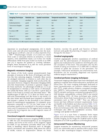
TABLE 16.9 A comparison of various imaging techniques for assessing brain structure haemodynamics
Imaging Technique Bedside use Spatial resolution Temporal resolution Scope of use Ease of interpretation
CEEG excellent good excellent excellent poor
Evoked potentials good fair Fair fair poor
Transcranial Doppler good fair Fair fair poor
MRI poor excellent poor good fair
Functional MRI poor excellent good poor poor
CT poor excellent poor good fair
Xenon CT poor good poor fair poor
ICP monitoring excellent poor good fair good
CEEG, continuous EEG; MRI, magnetic resonance imaging; CT, computed tomography; ICP, intracranial pressure.
important in neurological emergencies; (2) it clearly function, monitor the growth and function of brain
shows acute and sub-acute haemorrhages into the men- tumours and guide the planning of surgery or radiation
ingeal spaces and brain; and (3) it is less expensive than therapy for the brain. 49
45
a MRI. Disadvantages include: (1) it does not clearly
show acute or sub-acute infarcts or ischaemia, or brain Cerebral angiography
oedema, only established injury; (2) it does not clearly Cerebral angiography involves cannulation of cerebral
differentiate white from grey matter as clearly as an MRI; vessels and the administration of intraarterial contrast
and (3) it exposes the patient to ionising radiation. agents and medications for conditions involving the arte-
Despite these limitations it is still the most prevalent rial circulation of the brain. This procedure also has the
form of neurological imaging. 46 benefit of using non-invasive CT or MRI with or without
contrast to guide the accuracy of the procedure. For
Magnetic resonance imaging example, intracranial aneurysms and arteriovenous mal-
The tissues of the body contain proportionately large formations can be accurately diagnosed and repaired
amounts of protons in the form of hydrogen and func- without surgical intervention. 50
tion like tiny spinning magnets. Normally, these atoms Cerebral perfusion imaging techniques
are arranged randomly in relation to each other due to
the constantly changing magnetic field produced by the Numerous imaging techniques have been developed and
associated electrons. Magnetic Resonance Imaging (MRI) applied to evaluate brain haemodynamics, perfusion and
uses this characteristic of protons to generate images of blood flow. The main imaging techniques dedicated to
the brain and body. The advantages of MRI are: (1) it can brain haemodynamics are positron emission tomogra-
be manipulated to visualise a wide variety of abnormali- phy (PET), single photon emission computed tomogra-
ties within the brain; and (2) it can show a great deal of phy (SPECT), xenon-enhanced computed tomography
47
detail of the brain in normal and abnormal states. The (XeCT), dynamic perfusion computed tomography (PCT),
disadvantages of MRI are: (1) it does not show acute or MRI dynamic susceptibility contrast (DSC) and arterial
sub-acute haemorrhage into the brain in any detail; (2) spin labelling (ASL). All these techniques give similar
the time frame and enclosed space required to perform information about brain haemodynamics in the form of
51
and prepare a patient for the procedure is not advanta- parameters such as CBF or CBV. They use different
geous for neurological emergencies; (3) relatively more tracers and have different technical requirements. Some
expensive compared to CT; (4) the loud noise of the are feasible at the bedside and others not (see Table 16.9).
procedure needs to be considered in the patient manage- The duration of data acquisition and processing varies
ment; and (5) equipment for life support and monitoring from one technique to the other. Brain perfusion imaging
needs to be non-magnetic due to the magnetic nature of techniques also differ by quantitative accuracy, brain
the procedure. 48 coverage and spatial resolution. 52
Figure 16.13 is a scan from a traumatic brain injury
Functional magnetic resonance imaging patient and demonstrates a brain perfusion scan radio-
Functional magnetic resonance imaging (fMRI) is similar nuclide imaging. In the image the cerebral cortex is dark,
to MRI but uses deoxyhaemoglobin as an endogenous indicative of no CBF or perfusion confirming brain death.
contrast, and serves as the source of the magnetic signal
for fMRI. It can determine precisely which part of the Intracranial Pressure Monitoring
brain is handling critical functions such as thought, Invasive measures for monitoring intracranial pressure
speech, movement and sensation, help assess the effects (ICP) are commonly used in patients with a severe head
of stroke, trauma or degenerative disease on brain injury or after neurological surgery. Normal ICP varies