
Page 460 - ACCCN's Critical Care Nursing
P. 460
Neurological Assessment and Monitoring 437
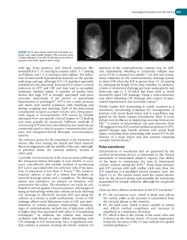
FIGURE 16.13 Brain death confirmed with brain per-
fusion scan radionuclide imaging. The cerebral cortex
is dark, indicative of no CBF. Permission received from
patient’s next of kin (patient brain dead).
with age, body position, and clinical condition. The insertion of the ventriculostomy catheter may be diffi-
normal ICP is 7–15 mmHg in a supine adult, 3–7 mmHg cult. Importantly, bleeding or ventricular collapse may
56
in children, and 1.5–6 mmHg in term infants. The defini- occur if CSF is drained too rapidly. For this last reason,
tion of intracranial hypertension depends on the specific many clinicians set the ventriculostomy drainage system
pathology and age, although ICP >15 mmHg is generally to drain CSF when the ICP is greater than 15–20 mmHg
considered to be abnormal. Increased ICP causes a critical by adjusting the height of the drip chamber. In addition,
reduction in CPP and CBF and may lead to secondary a limit of ventricular drainage per hour using gravity and
ischaemic cerebral injury. A number of studies have three-way taps to 5–10 mL/h has been used to avoid
shown that high ICP is strongly associated with poor excessively rapid CSF drainage. Using a ventriculostomy
outcome, particularly if the period of intracranial may allow lifesaving CSF drainage and control of intra-
53
hypertension is prolonged. ICP is not a static pressure cranial hypertension and secondary injury. 57
and varies with arterial pulsation, with breathing and Whilst routine ICP monitoring is widely accepted as a
during coughing and straining. Each of the intracranial mandatory monitoring technique for management of
constituents occupies a certain volume and, being essen- patients with severe head injury and is a guideline sug-
tially liquid, is incompressible. ICP cannot be reliably gested by the Brain Trauma Foundation, there is some
estimated from any specific clinical feature or CT finding debate over its efficacy in improving outcome from severe
and must actually be measured. Different methods of TBI. A review of neurocritical care and outcome from
58
monitoring ICP have been described but two methods are TBI suggested that ICP/cerebral perfusion pressure (CPP)-
commonly used in clinical practice: intraventricular cath- guided therapy may benefit patients with severe head
eters and intraparenchymal fibreoptic microtransducer injury, including those presenting with raised ICP in the
systems. absence of a mass lesion and also patients requiring
The reference point for the transducer is the foramina of complex interventions. 59
Monro (the duct joining the lateral and third ventricle
that is in alignment with the middle of the ear), although, Pulse waveforms
in practical terms, the external auditory meatus is Interpretation of waveforms that are generated by the
often used.
cerebral monitoring devices is important in the clinical
Currently, ventriculostomy is the most accurate (although assessment of intracranial adaptive capacity (the ability
the intraparenchymal fibreoptic is now similar in accu- of the brain to compensate for rises in intracranial
60
racy), cost-effective and reliable method of monitoring volume without raising the ICP). Brain tissue pressure
ICP and is associated with low infection risks if the dura- and ICP increase with each cardiac cycle and, thus, the
54
tion of placement is less than 72 hours. The ventricu- ICP waveform is a modified arterial pressure wave. See
lostomy catheter is part of a system that includes an Figure 16.14. The cardiac waves reach the cranial circula-
external drainage system and a transducer. The drainage tion via the choroid plexus and resemble the waveforms
system and transducer are primed on insertion with transmitted by arterial catheters, although the amplitude
preservative-free saline. The transducer can easily be cali- is lower.
brated or zeroed against a known pressure. Advantages of 61
using an indwelling ventricular catheter include allowing There are three distinct peaks seen in the ICP waveform:
CSF drainage to effectively decrease ICP and using the ● P1: the percussion wave, which is sharp and reflects
catheter as a means to instil medications. Access to CSF the cardiac pulse as the pressure is transmitted from
drainage allows serial laboratory tests of CSF and deter- the choroid plexus to the ventricle;
mination of volume–pressure relationships. Disadvan- ● P2: the tidal wave, which is more variable in nature
tages of ventriculostomy include risk of infection, which and reflects cerebral compliance and increases in
is higher than that associated with other ICP-monitoring amplitude as compliance decreases;
55
techniques. In addition, the catheter may become ● P3: which is due to the closure of the aortic valve and
occluded with blood or tissue debris, interfering with is known as the dicrotic notch. Of recent importance
CSF drainage or ICP monitoring. Also, if significant cere- is that the elevation of the P3 may indicate low global
bral oedema is present, locating the lateral ventricle for cerebral perfusion. 62