Page 473 - ACCCN's Critical Care Nursing
P. 473

450  P R I N C I P L E S   A N D   P R A C T I C E   O F   C R I T I C A L   C A R E

                                 ICP                              RAP < 0
                                                                  Deranged
                                                                  cerebrovascular
                                          RAP = 0       RAP = 1   reactivity
                                           Good           Poor
                                       compensatory   compensatory
                                          reserve        reserve
                          ‘Critical’ ICP                                          pulse amplitude  Pressure response-ICP


                                                                         Volume


                                               Pulsatile cerebral blood
                                                      volume              RAP = index of compensatory reserve.
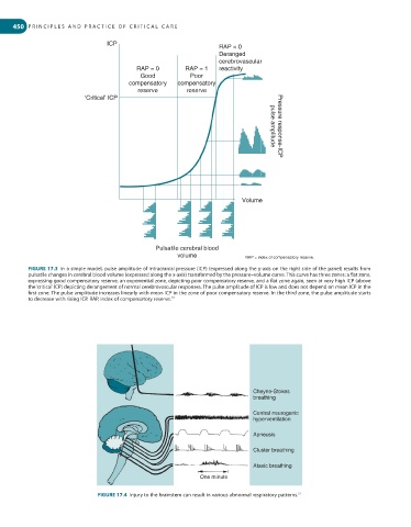
         FIGURE 17.3  In a simple model, pulse amplitude of intracranial pressure (ICP) (expressed along the y-axis on the right side of the panel) results from
         pulsatile changes in cerebral blood volume (expressed along the x-axis) transformed by the pressure–volume curve. This curve has three zones: a flat zone,
         expressing good compensatory reserve, an exponential zone, depicting poor compensatory reserve, and a flat zone again, seen at very high ICP (above
         the ‘critical’ ICP) depicting derangement of normal cerebrovascular responses. The pulse amplitude of ICP is low and does not depend on mean ICP in the
         first zone. The pulse amplitude increases linearly with mean ICP in the zone of poor compensatory reserve. In the third zone, the pulse amplitude starts
         to decrease with rising ICP. RAP, index of compensatory reserve.
                                                    26


                                                                            Cheyne-Stokes
                                                                            breathing

                                                                            Central neurogenic
                                                                            hyperventilation

                                                                            Apneusis

                                                                            Cluster breathing

                                                                            Ataxic breathing
                                                             One minute

                                                                                          27
                              FIGURE 17.4  Injury to the brainstem can result in various abnormal respiratory patterns.
   468   469   470   471   472   473   474   475   476   477   478