
Page 480 - ACCCN's Critical Care Nursing
P. 480
Neurological Alterations and Management 457
in Table 17.2 and is an adaptation of the current guide-
32
lines (see Table 17.3) to clinical practice (see Online
resources for TBI-related protocols). In all TBI multitrauma
patients, disability and exposure/environmental control
assessment includes the routine trauma series of X-rays,
namely chest, pelvis and cervical spine (lateral, anter-
oposterior and odontoid peg views). These should be
reviewed by a radiologist and areas of concern, parti-
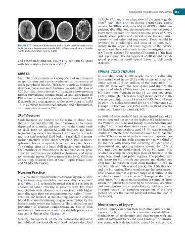
FIGURE 17.7 Extradural haematoma and a subtle subdural haematoma cularly in the upper and lower regions of the cervical
(left), subdural haematoma (middle left), diffuse axonal injury (middle spine, should be clarified with further investigations such
right), and combination injuries (right).
as CT scans. Isolated TBI requires CT scanning of the head
and upper spine. The management of TBI should include
and anterograde amnesia. Figure 17.7 contrasts CT scans spinal precautions until spinal injury is definitively
with haematoma formation and DAI. excluded.
SPINAL CORD TRAUMA
Mild TBI
In Australia, nearly 11,000 people live with a disability
Mild TBI often presents as a component of multitrauma from spinal cord injury (SCI), with an age-adjusted inci-
or sports injury and can be overlooked at the expense of dence rate of 13.6 per million of the population. In
75
other peripheral injuries. Risk factors such as vomiting, 2007–08 there were 362 new spinal cord injuries, the
dizziness, facial and skull fractures; including the loss of majority of which (79%) were due to traumatic causes.
CSF from the nose or the ear, will categorise those needing SCI were most frequent in the 15–24 year age group
further surveillance. Routine head CT and assessment of (30%), although trends show a significant increase in the
PTA are recommended to exclude mass lesions and DAI. average age at injury from 38 years in 1995–96 to 42 years
Diagnosis and management in the acute phase of mild in 2007–08. Males accounted for 84% of traumatic SCI.
TBI is as crucial to functional outcome and rehabilitation Transport-related injuries (46%) and falls (28%) were the
as in moderate-to-severe TBI. 72 main contributors to traumatic SCI.
Skull fractures In 2001–02 New Zealand had an unadjusted rate of 27
Skull fractures are present on CT scans in about two- per million and has one of the highest SCI incidences in
thirds of patients after TBI. Skull fractures can be linear, the Western world, related mostly to snowboarding and
60
depressed or diastatic, and may involve the cranial vault rugby. SCI occurs three times more often in men, and
or skull base. In depressed skull fractures the bone the incidence among those aged 15–34 years is roughly
fragment may cause a laceration of the dura mater, result- double the rate in those 35 years and over. More than half
ing in a cerebrospinal fluid leak. Basal skull fractures of the SCIs are due to vehicular trauma and a quarter due
73
include fractures of the cribriform plate, frontal bones, to motorcycle crashes. Falls account for nearly a third of
sphenoid bones, temporal bone and occipital bones. the injuries, with nearly half occurring in older people.
The clinical signs of a basal skull fracture may include: Recreational and sporting injuries account for 15% of
CSF otorrhoea or rhinorrhoea, haemotympanum, post- SCI, and 19% are work-related. Of all SCI cases, 51%
auricular ecchymoses, periorbital ecchymoses, and injury resulted in complete tetraplegia (loss of function in the
to the cranial nerves: VII (weakness of the face), VIII (loss arms, legs, trunk and pelvic organs). The predominant
of hearing), olfactory (loss of smell), optic (vision loss) risk factors for SCI include age, gender, and alcohol and
and VI (double vision). drug use. The vertebrae most often involved in SCI are
the 5th, 6th and 7th cervical (neck), the 12th thoracic,
and the 1st lumbar. These vertebrae are the most suscep-
Nursing Practice tible because there is a greater range of mobility in the
76
The surveillance and prevention of secondary injury is the vertebral column in these areas. Damage to the spinal
69
key to improving morbidity and mortality outcomes cord ranges from transient concussion or stunning (from
(see Table 17.1). It should be noted that in a post hoc in which the patient fully recovers) to contusion, laceration
analysis of saline critically ill patients with TBI, fluid and compression of the cord substance (either alone or
resuscitation with albumin was associated with higher in combination), to complete transection of the cord
74
mortality rates than was resuscitation with saline. Inter- (which renders the patient paralysed below the level of
ventions are targeted at maintaining adequate cerebral the injury).
blood flow and minimising oxygen consumption by the
brain in order to prevent ischaemia. The anticipation and Mechanisms of Injury
prevention of systemic complications are also of vital
importance. Assessment is vital to establish priorities in Cervical injury can occur from both blunt and penetrat-
care and is discussed in Chapter 16. ing trauma but in reality is a combination of different
mechanisms of acceleration and deceleration with and
77
Nursing management of the neurologically impaired, without rotational forces and axial loading. An illustra-
immobilised, mechanically ventilated patient is described tive example is a diving injury, caused by a direct load