
Page 479 - ACCCN's Critical Care Nursing
P. 479
456 P R I N C I P L E S A N D P R A C T I C E O F C R I T I C A L C A R E
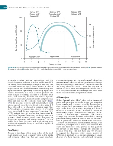
Defend CPP Optimise CPP Maintain CPP
Restore MAP Normalise MAP Maintain MAP
Reduce ICP Reduce ICP
120
30
Cerebral blood flow (mL/100 g/min) 40 C 10 Intracranial pressure (mmHg)
B
A
80
20
Hypoperfusion Hyperaemia Vasospasm
0
0 1 2 3 4 6 8 10 12 14
Days post-injury
FIGURE 17.6 Conceptual changes in cerebral blood flow and intracranial pressure (ICP) over time following traumatic brain injury: (A) cytotoxic oedema;
(B) vasogenic oedema; (C) cerebral blood flow CPP = cerebral perfusion pressure; MAP = mean arterial pressure.
ischaemia. Cerebral oedema, haemorrhage and bio- Contact phenomena are commonly superficial and can
chemical response to injury, infection and increased ICP generate superficial or contusional haemorrhages through
are among the commonest physiological responses that coup and contrecoup mechanisms. Cerebral contusions
71
can cause secondary injury. Tissue hypoxia is also of are readily identifiable on CT scans, but may not be
major concern and airway obstruction immediately after evident on day 1 scans, becoming visible only on days 2
injury contributes significantly to secondary injury. Poor or 3. Deep intracerebral haemorrhages can result from
cerebral blood flow, as a result of direct (primary) vas- either focal or diffuse damage to the arteries.
cular changes or damage, can lead to ischaemic brain
tissue, and eventually neuronal cell death. Systemic Diffuse injury
68
changes in temperature, haemodynamics and pulmonary
status can also lead to secondary brain injury (Figure Diffuse (axonal) injury (DAI) refers to the shearing of
17.6). In moderate to severe and, occasionally in mild, axons and supporting neuroglia; it may also traumatise
injury, cerebral blood flow is altered in the initial 2–3 blood vessels and can cause petechial haemorrhages,
71
days, followed by a rebound hyperaemic stage (days deep intracerebral haematomas and brain swelling.
4–7) leading to a precarious state (days 8–14) of cerebral DAI results from the shaking, shearing and inertial
vessel unpredictability and vasospasm. More than 30% effects of a traumatic impact. Mechanical damage to
64
of TBI patient have AN dysfunction characterised by small venules as part of the BBB can also trigger the for-
episodes of increased heart rate, respiratory rate, tem- mation of haemorrhagic contusions. This vascular
perature, blood pressure, muscle tone, decorticate or damage may increase neuronal vulnerability, causing
70
decerebrate posturing, and profuse sweating. Lack of post-traumatising perfusion deficits and the extravasa-
insight into these processes and implementing early tion of potentially neurotoxic blood-borne substances.
weaning of supportive therapies can lead to significant The most consistent effect of diffuse brain damage, even
secondary insults. when mild, is the presence of altered consciousness. The
depth and duration of coma provide the best guide to
the severity of the diffuse damage. The majority of
Focal injury patients with DAI will not have any CT evidence
Because of the shape of the inner surface of the skull, to support the diagnosis. Other clinical markers of
focal injuries are most commonly seen in the frontal DAI include the high speed or force strength of injury,
and temporal lobes, but they can occur anywhere. absence of a lucid interval, and prolonged retrograde