
Page 503 - ACCCN's Critical Care Nursing
P. 503
480 P R I N C I P L E S A N D P R A C T I C E O F C R I T I C A L C A R E
RELATED ANATOMY AND Organs and structures of the urinary system
PHYSIOLOGY
The renal system has a number of functions, including Adrenal
regulation and maintenance of fluid and electrolyte glands
balance, clearance of metabolic and other waste products, Right Kidney
an indirect role in the maintenance of blood pressure, renal
acid–base balance, and an endocrine function. In critical artery
care, an appreciation of the renal system’s fluid manage-
ment, blood pressure, electrolyte and acid–base functions
is essential.
Regulation and maintenance of the extracellular fluid and Right
electrolyte constituents is principally via the process of renal
filtration and reabsorption. The kidneys receive approxi- vein Aorta
mately 25% of the cardiac output each minute, and
excrete approximately 180 L/day of glomerular filtrate. Vena cava Ureter
Fortunately, tubular reabsorption accounts for approxi-
mately 178.5 L/day of the original filtrate, allowing for
a modest daily fluid intake of 1.5 L to achieve fluid
balance. During this process of filtration and reabsorp- Bladder
tion, metabolic byproducts, electrolyte and other wastes
(including many drugs) are also excreted and maintained
in balance. As with all body organ systems, an adequate
blood pressure and supply of oxygen to the kidneys is
paramount in maintaining the fluid and electrolyte
regulatory role.
ANATOMY OF KIDNEYS, NEPHRON AND
URINARY DRAINAGE SYSTEM
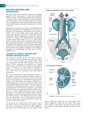
The functional anatomy of the renal system includes
the two kidneys, ureters, bladder and urethra (see Figure
18.1). The ureters, bladder and urethra collect, drain A Urethra
and temporarily store the urine produced from each
9
kidney. While important in providing the conduit
for the final excretion of urine, the kidney is the primary Frontal section of kidney
organ of interest in the renal system, particularly in
critical care practice, and hence will be described in Pyramid
more detail from the anatomical and physiological
perspectives. Papilla Fibrous
capsule
The kidneys are located in the retroperitoneal space on Minor
the posterior wall of the abdominal cavity, encased in a calyx
protective combination of the ribs, muscle, fat, tendon Renal
and the renal capsule. Each adult kidney weighs approxi- pelvis Major
mately 140 g. The kidneys may develop a different ana- calyx
tomical appearance, or vary in number and location from Medulla
the classic description provided here. The functional unit Ureter
of the kidney is the nephron, which consists of a filtrate- Cortex
collecting device (the Bowman’s capsule), a convoluted
tubule that varies in length and diameter, finally attach-
ing to a common filtrate-collecting tubule and duct
(see Figure 18.2). Within the Bowman’s capsule rests the B
glomerulus, a tuft of interlaced capillaries that arise from
the afferent arteriole. The efferent arteriole then drains FIGURE 18.1 Kidney and urinary drainage system.
107
from the glomerulus via a closely entwined network
called the peritubular capillaries, until these collect in the
venous network of the kidney.
kidney, which then drain into the renal pelvis where
The glomeruli and nephrons lie in the cortical area of urine is gathered to drain into the ureter. The major
the kidney, while the collecting ducts gather together blood vessels of the kidney, the renal artery and veins
into the renal pyramids, which lie in the medulla of also enter the renal capsule through the pelvis of the
the kidney. The pyramids drain into the calyces of the kidney. 9