
Page 657 - ACCCN's Critical Care Nursing
P. 657
634 S P E C I A LT Y P R A C T I C E I N C R I T I C A L C A R E
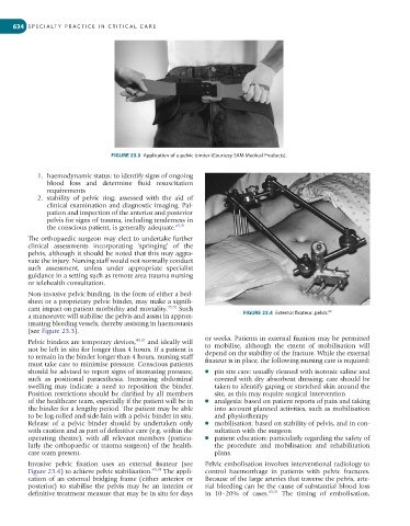
FIGURE 23.3 Application of a pelvic binder (Courtesy SAM Medical Products).
1. haemodynamic status: to identify signs of ongoing
blood loss and determine fluid resuscitation
requirements
2. stability of pelvic ring: assessed with the aid of
clinical examination and diagnostic imaging. Pal-
pation and inspection of the anterior and posterior
pelvis for signs of trauma, including tenderness in
the conscious patient, is generally adequate. 45,51
The orthopaedic surgeon may elect to undertake further
clinical assessments incorporating ‘springing’ of the
pelvis, although it should be noted that this may aggra-
vate the injury. Nursing staff would not normally conduct
such assessment, unless under appropriate specialist
guidance in a setting such as remote area trauma nursing
or telehealth consultation.
Non-invasive pelvic binding, in the form of either a bed-
sheet or a proprietary pelvic binder, may make a signifi-
cant impact on patient morbidity and mortality. 45,51 Such 80
a manoeuvre will stabilise the pelvis and assist in approx- FIGURE 23.4 External fixateur: pelvis.
imating bleeding vessels, thereby assisting in haemostasis
(see Figure 23.3).
or weeks. Patients in external fixation may be permitted
Pelvic binders are temporary devices, 45,51 and ideally will
not be left in situ for longer than 4 hours. If a patient is to mobilise, although the extent of mobilisation will
to remain in the binder longer than 4 hours, nursing staff depend on the stability of the fracture. While the external
must take care to minimise pressure. Conscious patients fixateur is in place, the following nursing care is required:
should be advised to report signs of increasing pressure, l pin site care: usually cleaned with isotonic saline and
such as positional paraesthesia. Increasing abdominal covered with dry absorbent dressing; care should be
swelling may indicate a need to reposition the binder. taken to identify gaping or stretched skin around the
Position restrictions should be clarified by all members site, as this may require surgical intervention
of the healthcare team, especially if the patient will be in l analgesia: based on patient reports of pain and taking
the binder for a lengthy period. The patient may be able into account planned activities, such as mobilisation
to be log-rolled and side-lain with a pelvic binder in situ. and physiotherapy
Release of a pelvic binder should by undertaken only l mobilisation: based on stability of pelvis, and in con-
with caution and as part of definitive care (e.g. within the sultation with the surgeon
operating theatre), with all relevant members (particu- l patient education: particularly regarding the safety of
larly the orthopaedic or trauma surgeon) of the health- the procedure and mobilisation and rehabilitation
care team present. plans.
Invasive pelvic fixation uses an external fixateur (see Pelvic embolisation involves interventional radiology to
Figure 23.4) to achieve pelvic stabilisation. 45,51 The appli- control haemorrhage in patients with pelvic fractures.
cation of an external bridging frame (either anterior or Because of the large arteries that traverse the pelvis, arte-
posterior) to stabilise the pelvis may be an interim or rial bleeding can be the cause of substantial blood loss
definitive treatment measure that may be in situ for days in 10–20% of cases. 45,51 The timing of embolisation,