
Page 718 - ACCCN's Critical Care Nursing
P. 718
Paediatric Considerations in Critical Care 695
Ventilation settings are reduced to minimal to minimise
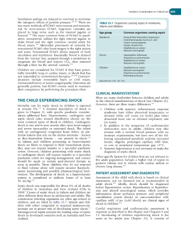
the iatrogenic effects of positive pressure. 156,157 There are TABLE 25.7 Organisms causing sepsis in newborns,
two main methods of ECMO: veno-venous and venoarte- infants and children
rial. In veno-venous ECMO, large-bore cannulas are
placed in large veins, such as the internal jugular or Age group Common organisms causing sepsis
femoral. 158 The more common form of ECMO in paedi-
atrics, venoarterial, utilises the right internal jugular to Newborns Group B beta-haemolytic streptococci
Enterobacteriaceae (such as E. coli)
drain blood and the right common carotid artery for Listeria monocytogenes
blood return. 158 Alternative placement of cannulas for Herpes simplex virus
venoarterial ECMO after heart surgery is the right atrium Staphylococcus aureus
and aorta. Venoarterial ECMO allows support of both Neisseria meningitidis
circulation and ventilation. Essentially, blood is drained Infants Haemophilus influenzae
from the ‘venous’ line, pumped through a membrane to Streptococcus pneumoniae
oxygenate the blood and remove CO 2 , then returned Staphylococcus aureus
Neisseria meningitidis
through a filter via the ‘arterial’ cannula. 158
Children Staphylococcus aureus
Children are considered for ECMO if they have poten- Neisseria meningitidis
tially reversible lung or cardiac injury, or shock that has Streptococcus pneumoniae
not responded to conventional therapies. 159-161 Contrain- Enterobacteriaceae
dications include irreversible brain or CNS injury, Adapted from (164, 165, 172).
immunodeficiency or severe coagulopathy. Outcomes are
generally positive, but ECMO centres need to maintain
their competence by performing the procedure often.
CLINICAL MANIFESTATIONS
There are many similarities between children and adults
THE CHILD EXPERIENCING SHOCK in the clinical manifestations of shock (see Chapter 21).
Mortality rate for septic shock in children is reported However, there are three major differences: 163
162
at around 9%. A detailed description of shock is 1. Children with systemic inflammatory response
given in Chapter 21, with specific paediatric consider- syndrome have either abnormal temperature or
ations addressed here. Hypovolaemic, cardiogenic and elevated white cell count (or both) plus either
septic shock (also termed distributive shock) are the abnormal heart rate or elevated respiratory rate
most common types of shock in children. Cardiogenic (or both).
shock is rare and is seen mainly after open-heart surgery 2. In addition to the symptoms of cardiovascular
and severe myocarditis or untreated shock. The infant dysfunction seen in adults, children may also
with an undiagnosed congenital heart defect, in par- present with a normal blood pressure with no
ticular lesions that rely on the ductus arteriosis – known inotrope requirements, but have two of the fol-
as duct-dependent lesions – can present in shock. 162 lowing: unexplained metabolic acidosis, increased
As infants and children presenting in hypovolaemic lactate, oliguria, prolonged capillary refill time,
shock are likely to respond to fluid resuscitation alone, or core to peripheral temperature gap >3°C.
they may not require transfer to a specialist paediatric 3. Systemic hypotension is not necessary to make the
centre. However, children presenting with septic shock diagnosis of septic shock.
or cardiogenic shock will require transfer to a specialist
paediatric centre for ongoing management, and contact Other specific factors for children that are not relevant in
should be made to initiate goal-directed therapy as the adult population include a higher risk of sepsis in
soon as possible. Those children who do not respond preterm infants and in infants with cardiac defects or
162
to fluid volume alone will require invasive haemody- chronic lung disease.
namic monitoring and possible pharmacological inter-
vention. The development of shock in a hypovolaemic PATIENT ASSESSMENT AND DIAGNOSTIC
patient is considered to indicate losses of at least Assessment of the child with shock is based on clinical
30 mL/kg. 162 assessment, not on chemical test as recommended in
adult shock. 162 Ideally, shock should be diagnosed
Septic shock was responsible for about 8% of all deaths before hypotension occurs. Hypothermia or hyperther-
of children in Australian and New Zealand ICUs in mia and altered neurological status, which provides
4
2008. Causes of septic shock in infants and children are information about perfusion pressure and peripheral
often different from those in adolescents and adults. The vasodilation (warm shock) or vasoconstriction with
commonest infecting organisms are often age-related in capillary refill >2 sec (cold shock) are clinical signs of
children, and are listed in Table 25.7. Infants and chil- shock in children. 162
dren with either congenital or acquired immunocom-
16
promise are at greater risk of developing septic shock. Careful respiratory and cardiovascular assessment is
Meningococcal sepsis remains the leading cause of septic required, as described in this chapter and Chapters 9 and
shock in developed countries such as Australia and New 13. Monitoring of children experiencing shock is the
Zealand. same as for adults (see Chapter 21). It consists of