


Page 115 - Concise Pathology for Exam Preparation ( PDFDrive )
P. 115
100   SECTION I    General Pathology


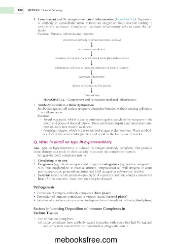
                        	�Complement and Fc receptor-mediated inflammation (Flowchart 5.6): Deposition
                     3 .

                        of    antibody      extracellular  tissue  initiates  an  antigen-antibody  reaction  leading  to


                                  i

                                   n

                        complement    activation.  Complement  activates  inflammatory  cells      cause  the  cell


                                                                               t

                                                                                o

                        injury.
                        Examples:   Parasitic infections and tumours
                                       Deposition of antibody in extracellular tissue, eg, ECM
                                                Activation of complement
                                  Generation of C3a and C5a, which recruit neutrophils and monocytes
                                  Inflammatory cells bind to deposited antibodies via their Fc receptors
                                                 Activation of leukocytes
                                             Release of enzymes and free radicals
                                                    Tissue damage
                            FLOWCHART 5.6.    Complement and Fc receptor-mediated inflammation.
                     4.
                        	�Antibody-mediated cellular dysfunction
                        Antibodies against cell surface receptors deregulate function without causing cell injury
                          or inflammation.
                        Examples

                            •	�Myasthenia gravis, which is due to antibodies against acetylcholine receptors in the
                           motor end plates of skeletal muscle. These antibodies impair neuromuscular trans-
                           mission and cause muscle weakness.

                            •	�Pemphigus vulgaris, which is due to antibodies against desmosomes. These antibod-
                           ies disrupt the intercellular junction and result in the formation of vesicles.
                     Q.   Write in detail on type III hypersensitivity.
                     Ans.   Type III hypersensitivity is induced by antigen–antibody complexes that produce
                     tissue damage as a result of their capacity to activate the complement system.
                        Antigen–antibody complexes may be:
                        	�Circulating or in situ

                     1.
                     2.
                        	�Exogenous (eg, infectious agents and drugs) or endogenous (eg, ‘nuclear antigens’ in


                        SLE, ‘immunoglobulins’ in reactive arthritis, ‘streptococcal cell wall antigens’ in acute
                        post-streptococcal glomerulonephritis and ‘HBS antigen’   in polyarteritis nodosa)

                     3.
                        	�Systemic (acute serum sickness—prototype of a systemic immune complex disease) or
                        local   (Arthus reaction—local immune complex disease)
                     Pathogenesis
                        •	�Formation of antigen–antibody complexes (first phase)
                        •	�Deposition of immune complexes in various tissues (second phase)


                        •	�Initiation of an inflammatory reaction in dispersed sites throughout the body (third phase)


                     Factors Influencing Deposition of Immune Complexes in
                     Various Tissues
                     1.
                         Size of immune complexes:
                        (a)
                            Large complexes have antibody excess (complex with many free IgG Fc regions)
                           and are readily removed by the mononuclear phagocytic system.
                                  mebooksfree.com

