


Page 111 - Concise Pathology for Exam Preparation ( PDFDrive )
P. 111
96 SECTION I General Pathology
Antigenpresenting cell
MHC CD80 (B7–1)
or
CD86 (B7–2)
Ag 2
CD4
or V
CD8 TCR
C
CD28
1
CD3 proteins T cell
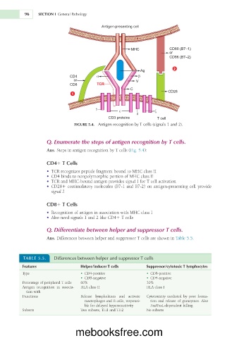
FIGURE 5.4. Antigen recognition by T cells (signals 1 and 2).
Q. Enumerate the steps of antigen recognition by T cells.
Ans. Steps in antigen recognition by T cells (Fig. 5.4):
CD41 T Cells
• TCR recognizes peptide fragment bound to MHC class II
• CD4 binds to nonpolymorphic portion of MHC class II
• TCR and MHC-bound antigen provides signal 1 for T cell activation
• CD281 costimulatory molecules (B7-1 and B7-2) on antigen-presenting cell provide
signal 2
CD81 T Cells
• Recognition of antigen in association with MHC class I
• Also need signals 1 and 2 like CD41 T cells
Q. Differentiate between helper and suppressor T cells.
Ans. Differences between helper and suppressor T cells are shown in Table 5.5.
TABLE 5.5. Differences between helper and suppressor T cells
Features Helper/inducer T cells Suppressor/cytotoxic T lymphocytes
Type • CD4-positive • CD8-positive
• CD8-negative • CD4-negative
Percentage of peripheral T cells 60% 30%
Antigen recognition in associa- HLA class II HLA class I
tion with
Functions Release lymphokines and activate Cytotoxicity mediated by pore forma-
macrophages and B cells, responsi- tion and release of granzymes. Also
ble for delayed hypersensitivity Fas/FasL-dependent killing
Subsets Two subsets, TH1 and TH2 No subsets
mebooksfree.com

