


Page 114 - Concise Pathology for Exam Preparation ( PDFDrive )
P. 114
5 Diseases of Immunity 99
TNFα Epithelial cells
Release of:
• Eotaxin
• RANTES (regulated on activation,
normal T expressed and secreted)
Recruitment of eosinophils
• Produce leukotriene C4 and PAF Contain:
• Activate mast cells • Major basic protein
• Eosinophilic cationic protein
Promote inflammation Toxic to epithelial cells
FLOWCHART 5.3. Role of eosinophils in type I hypersensitivity.
Q. Write in detail on type II hypersensitivity.
Ans. Type II hypersensitivity is mediated by antibodies directed towards antigens present
on the surface of the cells or other tissue components. These antigens may be intrinsic to
cell membrane or exogenous antigens absorbed on the cell surface (eg, a drug metabolite).
Reaction occurs when antibodies bind to normal or altered cell surface antigens.
Mechanisms Underlying Type II Hypersensitivity
1.
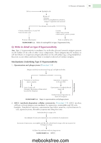
�Opsonization and phagocytosis (Flowchart 5.4)
Antigen–antibody reactions involving IgG and IgM antibodies
Complement activation
Formation of membrane Generation of opsonins
Direct lysis
attack complex
(C3b and C4b)
Lysis of target cell
target cell
(Example: destruction of thinwalled Opsonization of antibodybound Opsonization
bacteria like Neisseria)
Destruction by phagocytes via
their C3b receptors
FLOWCHART 5.4. Steps in opsonization and phagocytosis.
2.
�ADCC (antibody-dependent cellular cytotoxicity; Flowchart 5.5): ADCC involves
cell lysis without phagocytosis mediated by monocytes, neutrophils and NK cells.
Examples: Transfusion reactions, autoimmune haemolytic anaemia, erythroblastosis fe-
talis, agranulocytosis, thrombocytopenia and drug reactions
Antibodies bind to target cells
Recruitment of leukocytes (minimum or no activation of complement)
Activation of monocytes, neutrophils and NK cells, which bind to target cells via receptors for
Fc fragment of IgG
Cell lysis (by perforins) without phagocytosis
FLOWCHART 5.5. ADCC.
mebooksfree.com

