


Page 131 - Concise Pathology for Exam Preparation ( PDFDrive )
P. 131
116   SECTION I    General Pathology

                     Staining Characteristics

                        •	�Congo red
                          •	�Ordinary light-pink or red colour
                          •	�Polarized light-apple green birefringence (due to cross-b-pleated configuration)


                        •	�Metachromatic stains (Rosaniline dyes): Examples are methyl violet and crystal violet.


                       Amyloid   takes up a rose pink colour with these dyes.


                        •	�Fluorescent stains of Thioflavin T and S: In ultraviolet light, amyloid fluoresces yellow.


                        •	�Immunohistochemistry: Anti-AA and anti-lambda, anti-kappa antibodies can be used

                       to differentiate between different types of amyloid.

                        •	�Toluidine blue: Blue colour in ordinary light and dark red, birefringence under polar-
                       ized microscopy.
                        •	�Alcian blue: Blue-green colour.

                        •	�PAS (periodic acid-Schiff) and H&E stains: Pink colour.

                     Histopathology
                     Kidneys   (most common and most serious form of organ involvement)
                     Gross features
                        •	�Kidneys are normal or enlarged in early stage and shrunken or contracted in late stage
                       (amyloid deposition causes vascular narrowing leading to shrinking of the organ)
                     Microscopic features
                        •	�Primarily glomerular deposits
                        •	�Subtle thickening of mesangial matrix due to mesangial deposits
                        •	�Uneven widening of basement membrane of glomerular capillaries leading to capillary
                       narrowing due to basement membrane deposits
                        •	�Distortion of glomerular vascular tuft due to confluent masses of or interlacing broad
                       ribbons of amyloid
                     Spleen
                     Gross features:
                     Moderate to marked splenomegaly
                     Microscopic features:
                     There are two patterns of deposition -
                     1.
                         Sago  spleen:  Deposits  largely  limited  to  splenic  follicles;  entire  follicle  replaced  by
                        amyloid, leading to Tapioca-like granules
                     2.
                         Lardaceous spleen: Sparing of follicles; involvement of walls of splenic sinuses and con-
                        nective tissue framework of red pulp, leading to large map like areas
                     Liver
                     Gross features:
                     Moderate to marked hepatomegaly
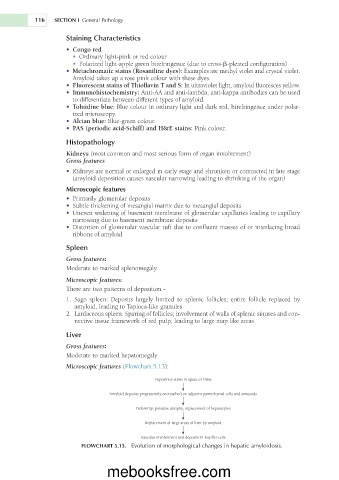
                     Microscopic features   (Flowchart 5.13):
                                                 Deposition starts in space of Disse
                                   Amyloid deposits progressively encroached on adjacent parenchymal cells and sinusoids
                                           Deformity, pressure atrophy, replacement of hepatocytes
                                              Replacement of large areas of liver by amyloid

                                            Vascular involvement and deposits in Kupffer cells
                          FLOWCHART 5.13.    Evolution of morphological changes in hepatic amyloidosis.


                                  mebooksfree.com

