


Page 143 - Concise Pathology for Exam Preparation ( PDFDrive )
P. 143
128   SECTION I  General Pathology


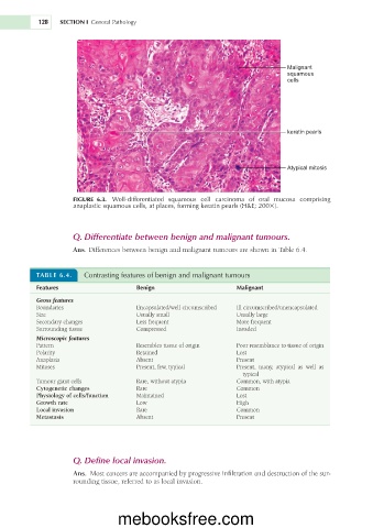
                                                                                 Malignant
                                                                                 squamous
                                                                                 cells


                                                                                 keratin pearls


                                                                                 Atypical mitosis


                      FIGURE  6.3.  Well-differentiated  squamous  cell  carcinoma  of  oral  mucosa  comprising
                      anaplastic squamous cells, at places, forming keratin pearls (H&E; 2003).


                     Q. Differentiate between benign and malignant tumours.

                     Ans. Differences between benign and malignant tumours are shown in Table 6.4.


           TABLE 6.4.    Contrasting features of benign and malignant tumours

           Features                    Benign                      Malignant
           Gross features
           Boundaries                  Encapsulated/well circumscribed  Ill circumscribed/unencapsulated
           Size                        Usually small               Usually large
           Secondary changes           Less frequent               More frequent
           Surrounding tissue          Compressed                  Invaded
           Microscopic features
           Pattern                     Resembles tissue of origin  Poor resemblance to tissue of origin
           Polarity                    Retained                    Lost
           Anaplasia                   Absent                      Present
           Mitoses                     Present, few, typical       Present,  many,  atypical  as  well  as
                                                                     typical
           Tumour giant cells          Rare, without atypia        Common, with atypia
           Cytogenetic changes         Rare                        Common
           Physiology of cells/function   Maintained               Lost
           Growth rate                 Low                         High
           Local invasion              Rare                        Common
           Metastasis                  Absent                      Present


                     Q. Define local invasion.
                     Ans.  Most cancers are accompanied by progressive infiltration and destruction of the sur-
                     rounding tissue, referred to as local invasion.


                                  mebooksfree.com

