


Page 141 - Concise Pathology for Exam Preparation ( PDFDrive )
P. 141
126   SECTION I  General Pathology

                     Q. Define anaplasia. Enumerate the morphological features indicative
                     of anaplasia.
                     Ans.  Anaplasia is defined as lack of differentiation; literal meaning is ‘reverse differentia-
                     tion’. It is a hallmark of malignant transformation.

                     Morphological Features Indicative of Anaplasia
                     •  Pleomorphism (variation in size and shape of cells)
                     •  Anisonucleosis (variation in size of nuclei)
                     •  Abnormal nuclear morphology:
                       •  Abundant and darkly staining (hyperchromatic) DNA
                       •  Coarsely clumped chromatin or clumping of nuclear chromatin along the nuclear
                         membrane resulting in prominent appearing nucleoli
                       •  Increased nucleocytoplasmic ratio (normal from 1:4 to 1:6; may approach 1:1)
                     •  Numerous mitoses with abnormal, atypical, bizarre, tri, quadri and multipolar spindles
                       (abnormal mitoses are seen in malignant tumours only)
                     •  Loss of polarity (orientation of cells)
                     •  Presence of the tumour giant cells

                     Q. Define dysplasia. Enumerate the steps in the course of progression
                     of dysplasia to invasive cancer.
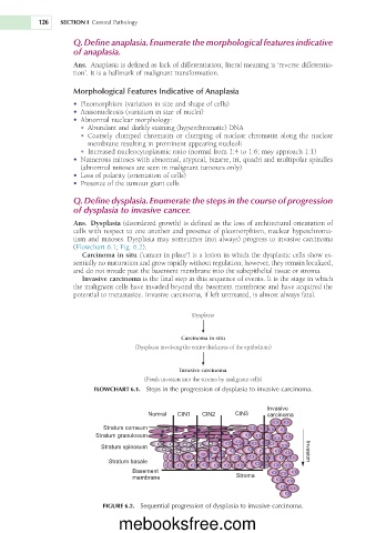
                     Ans.  Dysplasia (disordered growth) is defined as the loss of architectural orientation of
                     cells with respect to one another and presence of pleomorphism, nuclear hyperchroma-
                     tism and mitoses. Dysplasia may sometimes (not always) progress to invasive carcinoma
                     (Flowchart 6.1; Fig. 6.2).
                        Carcinoma in situ (‘cancer in place’) is a lesion in which the dysplastic cells show es-
                     sentially no maturation and grow rapidly without regulation; however, they remain localized,
                     and do not invade past the basement membrane into the subepithelial tissue or stroma.
                        Invasive carcinoma is the final step in this sequence of events. It is the stage in which
                     the malignant cells have invaded beyond the basement membrane and have acquired the
                     potential to metastasize. Invasive carcinoma, if left untreated, is almost always fatal.


                                                      Dysplasia


                                                   Carcinoma in situ
                                      (Dysplasia involving the entire thickness of the epithelium)


                                                   Invasive carcinoma
                                         (Frank invasion into the stroma by malignant cells)
                           FLOWCHART 6.1.  Steps in the progression of dysplasia to invasive carcinoma.

                                                                           Invasive
                                          Normal  CIN1   CIN2     CIN3     carcinoma

                              Stratum corneum
                            Stratum granulosum
                             Stratum spinosum                                         Invasion

                               Stratum basale
                                      Basement
                                      membrane                     Stroma


                              FIGURE 6.2.  Sequential progression of dysplasia to invasive carcinoma.

                                  mebooksfree.com

