


Page 144 - Concise Pathology for Exam Preparation ( PDFDrive )
P. 144
6  Neoplasia  129

             Q. Define metastasis. Write briefly on the various pathways of spread
             of the tumours.
             Ans.  Tumour implants discontinuous with the primary tumour, confirm the malignant
             nature of a tumour and are labelled metastases. All cancers metastasize with a few excep-
             tions, eg, basal cell carcinoma (rodent ulcer) and gliomas of the central nervous system,
             which are locally invasive and rarely metastasize.

             Pathways of Spread of the Tumours
               1.  Direct seeding of body cavities and surfaces: Penetration of a tumour into a natural
                open field/space, eg, pleural, pericardial, subarachnoid and synovial. Sometimes muci-
                nous tumours of appendix and ovary (both benign and malignant) fill the peritoneal
                cavity with a gelatinous neoplastic mass called ‘pseudomyxoma peritonei’.
               2.  Lymphatic spread
                 (a)  There are numerous interconnections between lymphatic and vascular channels;
                   so,  emphasis  on  differentiating  lymphatic  and  vascular  dissemination  may  be
                   purposeless.
                 (b)  Functional lymphatics are absent in tumours and lymphatic vessels located at the
                   surface are sufficient for lymphatic spread.
                 (c)  Lymphatic spread tends to follow natural routes of lymphatic drainage and is the
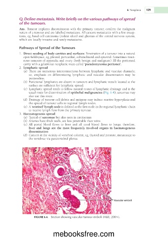
                   usual route for dissemination of epithelial malignancies (Fig. 6.4); sarcomas may
                   also use this route.
                  (d)  Drainage of tumour cell debris and antigens may induce reactive hyperplasia and
                   the spread of tumour cells to regional lymph nodes.
                 (e)  A ‘sentinel’ lymph node is defined as the first node in the regional lymphatic chain
                   to receive lymph flow from the primary tumour.
               3.  Haematogenous spread
                 (a)  Typical of sarcomas but also seen in carcinomas
                 (b)  Arteries have thick walls, are less penetrable than veins
                 (c)  All  portal  blood  flows  to  liver  and  all  caval  blood  flows  to  lungs;  therefore,
                   liver and lungs are the most frequently involved organs in haematogenous
                   dissemination
                  (d)  Cancers in the vicinity of vertebral column, eg, thyroid and prostate, metastasize to
                   the vertebrae via paravertebral plexus


                                                                        Vascular emboli


                      FIGURE 6.4.  Section showing vascular tumour emboli (H&E; 2003).


                                  mebooksfree.com

