


Page 149 - Concise Pathology for Exam Preparation ( PDFDrive )
P. 149
134   SECTION I  General Pathology

                     Q. Differentiate between anti-oncogenes and proto-oncogenes.

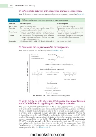
                     Ans.  Differences between anti-oncogenes and proto-oncogenes are enlisted in Table 6.6.


           TABLE 6.6.    Differences between anti-oncogenes and proto-oncogenes
           Features    Anti-oncogenes                          Proto-oncogenes
           Other name  Tumour suppressor genes                 Precursor genes for oncogene
           Function    They suppress cell proliferation, and promote differ-  They promote normal cell growth and dif-
                         entiation and maturation of cells      ferentiation
           Inheritance  Recessive. Homozygous inactivation, ie, loss of both   Dominant. Mutation in a single copy may
                         normal copies of gene is required for carcinogenesis  lead to oncogenic conversion
           Action      Act  passively,  ie,  cancer-promoting  genes  dominate   Act actively, ie, gene products of oncogenes
                         and lead to tumour formation due to loss of normal   directly lead to tumour formation
                         function of anti-oncogenes
           Examples    P53, RB gene, BRCA-1 and 2, TGF-b, APC, WT-1 and NF  myc, N-myc, erbB1/2/3


                     Q. Enumerate the steps involved in carcinogenesis.
                     Ans.  Carcinogenesis is a multistep process (Flowchart 6.2).

                                   Acquired or       Normal cell
                                   environmental                DNA repair
                                   DNA-damaging agents
                                                     DNA damage
                                INITIATION OF CELL  Activation of growth-  Failure of DNA repair   Inherited mutations


                                             Mutations in the genome of somatic cells
                                                                      Abnormalities in genes
                                                   Inactivation of tumour
                                                    suppressor genes
                                    promoting genes
                                                                       regulating apoptosis
                                                                       Decreased apoptosis
                                PROGRESSION        Malignant neoplasm
                                                    Clonal expansion


                                                 Angiogenesis  New mutations
                                                   Invasion/metastasis
                                     FLOWCHART 6.2.  Steps involved in carcinogenesis.

                     Q. Write briefly on role of cyclins, CDK (cyclin-dependent kinases)
                     and CDK inhibitors in regulating G 1 /S cell cycle transition.
                     Ans. Cyclin D, the first cyclin to increase in the cell cycle, appears in mid-G phase, but is
                     no longer detectable in the S phase. There are three forms of cyclin D, named Dl, D2 and
                     D3; but to simplify matters, the general term ‘cyclin D’ is used. Cyclin D, like other cyclins,
                     is  unstable  and  is  degraded  through  ubiquitin—proteasome  pathway.  During  the
                     G phase of the cell cycle, cyclin D binds to and activates CDK4, forming a cyclin D–CDK4
                     complex. This complex plays a critical role in the cell cycle by phosphorylating the retino-
                     blastoma  susceptibility  protein  (pRB).  ‘The  phosphorylation  of  RB  is  a  molecular
                     ON–OFF switch for the cell cycle.’ In its hypophosphorylated state, RB prevents cells
                     from replicating by forming a tight, inactive complex with the transcription factor E2F.
                     Phosphorylation  of  RB  dissociates  the  complex  and  releases  the  inhibition  on  E2F
                     transcriptional activity (see Flowchart 6.3).


                                  mebooksfree.com

