


Page 153 - Concise Pathology for Exam Preparation ( PDFDrive )
P. 153
138   SECTION I  General Pathology


                        •  He suggested that in hereditary cases one genetic change (‘first hit’) is inherited
                          from an affected parent, and is therefore present in all somatic cells of the body;
                          whereas, the second mutation (‘second hit’) occurs in one of the many retinal cells
                          (which already carry the first mutation).
                        •  In sporadic  cases, however, the two mutations (hits) occur somatically within a
                          single retinal cell whose progeny then form the tumour.
                        •  The mutations required to produce retinoblastoma involve the RB gene, located on
                          chromosome 13ql4.
                        •  Both normal alleles of the RB locus must be inactivated (two hits) for the develop-
                          ment of retinoblastoma.
                        •  In familial cases, children are born with one normal and one defective copy of the
                          RB gene. They lose the intact copy in the retinoblasts through some form of somatic
                          mutation (point mutation, deletion of 13q 14 or even complete loss of the normal
                          chromosome 13).
                        •  In sporadic cases, both normal RB alleles are lost by somatic mutation in one of the
                          retinoblasts.
                        •  Patients with familial retinoblastoma are also at a greatly increased risk of developing
                          osteosarcoma and some other soft-tissue sarcomas.
                        •  Furthermore, inactivation of the RB locus has been noted in several other tumours, includ-
                          ing adenocarcinoma of the breast, small cell carcinoma of the lung and bladder carcinoma.
                        •  Most importantly, alterations in the key regulators of the cell cycle or RB pathway
                          (involving INK4a proteins, cyclin D-dependent kinases and RB family proteins) are
                          present in most cancer cells. The RB gene can loose its normal suppressor action and
                          go into the proliferative mode by various mechanisms (loss of function mutations af-
                          fecting RB; gene amplification of CDK4 and cyclin D; loss of cyclin-dependant kinase
                          inhibitors like p16/INK4 and inhibition of RB by binding of viral oncoproteins to it).
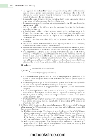
                        RB pathway:

                                    Active RB gene (hypophosphorylated)
                               S
                        G 1
                                    Inactive RB gene (hyperphosphorylated)
                        •  The retinoblastoma gene encodes a 110-kDa phosphoprotein (pRB) that is ex-
                          pressed in almost every cell of the human body and contributes to growth regulation
                          in these cells.
                        •  The most important target of the retinoblastoma protein is cellular transcription fac-
                          tor E2F. E2F is a potent stimulator of cell cycle entry into S phase.
                        •  E2F activity consists of an E2F polypeptide and a DP protein (E2F/DP heterodimeric
                          complex).
                        •  In the hypophosphorylated form (which occurs early in G 1 ), RB binds to E2F, inac-
                          tivates E2F as a transcription factor and shuts the expression of E2F target genes off.
                        •  Phosphorylation of pRB by Cyclin/CDK complexes in mid-to-late G 1  causes pRB to
                          lose its affinity for E2F.
                        •  The free E2F/DP transcription factor can now activate transcription of E2F target genes.
                        •  In  a  normal  cell  during  mitosis,  phosphatase  1-like  protein  (PP1)  removes  all
                          phosphates from pRB.
                       2.  Tp53 gene (p53): Situated on short arm of chromosome 17, it is also called ‘protector
                        of the genome’ as P53 mutation is present in more than half of all human cancers. P53
                        has two major functions:
                         (a)  Blocking mitotic activity: DNA damage is sensed by kinases of the ATM/ATR (ataxia
                           telangiectasia mutated or ataxia telangiectasia and Rad3) family which phosphory-
                           late p53, releasing it from inhibitors like MDM2 (mouse double minute 2 homolog).
                           If the damage is minor, activated p53 enhances expression of CDK inhibitor p21
                           which arrests/halts the cell-cycle at the G 1 /S checkpoint until the damage is repaired.
                         (b)  Role in promoting apoptosis: If the damage is major and cannot be repaired, p53
                           triggers the cell to commit suicide by apoptosis (activates apoptosis, inducing BAX
                           gene to bring the defective cell to an end).


                                  mebooksfree.com

