


Page 162 - Concise Pathology for Exam Preparation ( PDFDrive )
P. 162
6  Neoplasia  147


                   Air-dried  MGG  (May–Grünwald–Giemsa)-stained  smears  or  wet  (95%  ethanol)-
                     fixed–Haematoxylin-and-Eosin  stained  smears  are  prepared  from  the  FNAC
                     material obtained and examined.
                 (b)  Exfoliative cytology: Due to loss of cohesiveness, neoplastic cells are continuously
                   shed from tumours into the surrounding space. These are called exfoliated cells
                   and can be examined in the following preparations:
                     (i)  Papanicolaou smears (for carcinoma cervix)
                    (ii)  Sputum and bronchial washings (for bronchogenic carcinoma)
                     (iii)  Pleural, pericardial and peritoneal fluid (for local cancers)
                    (iv)  Urine (for urothelial malignancies)
                    (v)  Cerebrospinal fluid (for CNS tumours)
                    (vi)  Gastric secretions (for gastric carcinoma)
                       Diagnostic reliability of exfoliative cytology varies between 80% and 97%.
               2.  Histopathology: Histopathological diagnosis entails microscopy supported by clinical
                and  investigative  data.  Formalin  fixation  is  required  for  routine  histopathology  and
                glutaraldehyde fixation is required for electron microscopy. Frozen sectioning aids in
                rapid/intraoperative diagnosis.
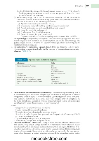
               3.  Histochemistry/cytochemistry (special stains): These are diagnostic tools for identi-
                fying chemical composition of cells for the purpose of tumour diagnosis and clas-
                sification (Table 6.9).


                   TABLE 6.9.   Special stains in tumour diagnosis

                   Substances                          Stain
                   Basement membrane/collagen          PAS
                                                       Reticulin
                                                       Van Gieson
                                                       Masson’s trichrome
                   Glycogen                            PAS with diastase loss
                   Glycoproteins                       PAS
                   Mucins of epithelial origin         Mucicarmine
                   Acid mucins (of mesenchymal origin)  Alcian blue
                   Argyrophilic/argentaffin granules/fungi  Silver stains
                   Fat                                 Oil red-O, Sudan black B


               4.  Immunohistochemistry/immunocytochemistry:  Immunohistocytochemistry  (IHC)
                is  an  immunological  method  of  recognizing  a  cell  based  on  recognition  of  specific
                components called ‘antigens’. ‘Specific antibodies’ against antigens are raised by hybrid-
                oma technique and labelled monoclonal antibodies. Antigen–antibody complexes on
                the slides (histological sections or cytology smears) are made visible for microscopic
                identification by labels (fluorochromes or  enzyme systems).
             Uses
               •  Categorization of undifferentiated neoplasms
               •  Specific typing of leukaemias/lymphomas
               •  Determination of site of origin of a metastatic tumour
               •  Detection  of  molecules  that  have  prognostic  or  therapeutic  significance,  eg,  ER–PR
                 receptors in carcinoma breast
               •  Expression of protein products of oncogenes
               •  Differentiating benign from malignant lesions
              5.  Intermediate filaments (IFs): IFs are a family of related proteins that share common struc-
                tural features. They have an average diameter of 10 nanometers, which is between that of
                microfilaments (which are smaller) and microtubules (which are larger). Most types of in-
                termediate filaments are cytoplasmic except lamins, which are nuclear. The most important
                function of intermediate filaments is to provide mechanical support for the plasma mem-
                brane, where they come in contact with other cells or with the extracellular matrix. Unlike


                                  mebooksfree.com

