


Page 20 - Concise Pathology for Exam Preparation ( PDFDrive )
P. 20
1  Cell Injury and Cell Death  5


             Hyperplasia
             Definition
             Increase in number of cells in an organ or tissue leading to increased size/mass of the tissue
             or organ. Hyperplasia takes place in cells, which are capable of synthesizing DNA. In nondivid-
             ing cells, only hypertrophy occurs.
             Mechanism
             •  Production of transcription factors that induce genes encoding growth factors, receptors
               for growth factors and cell-cycle regulators.
             •  In hormonal hyperplasia, hormones themselves act as growth factors and trigger tran-
               scription of genes.
             •  In compensatory hyperplasia, there is proliferation of remaining cells and development of
               new cells from stem cells.
             Types
               1.  Physiologic hyperplasia:
                 (a)  Hormonal hyperplasia: Hormonal stimulation increases the functional capacity of
                   the tissue when needed, eg, breast and uterus in puberty, pregnancy and lactation.
                 (b)  Compensatory hyperplasia: Increase in tissue mass after damage or partial resection,
                   eg, regeneration of liver after partial hepatectomy.
               2.  Pathologic hyperplasia: Hyperplasia due to excessive hormonal stimulation or excessive
                effects of growth factors on target cells, eg, endometrial hyperplasia (occurs when bal-
                ance  between  progesterone  and  oestrogen  is  disturbed)  and  benign  nodular  prostatic
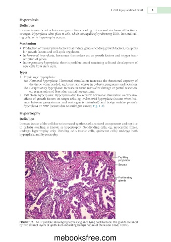
                hyperplasia or NHP (occurs due to androgen excess; Fig. 1.2).
             Hypertrophy

             Definition
             Increase in size of the cell due to increased synthesis of structural components and not due
             to  cellular  swelling  is  known  as  hypertrophy.  Nondividing  cells,  eg,  myocardial  fibres,
             undergo  hypertrophy  only.  Dividing  cells  (stable  cells,  quiescent  cells)  undergo  both
             hyperplasia and hypertrophy.


                                                                         Papillary
                                                                         projection
                                                                         Stroma


                                                                         Proliferating
                                                                         glands


             FIGURE 1.2.  NHP prostate showing hyperplastic glands lying back to back. The glands are lined
             by two distinct layers of epithelium indicating benign nature of the lesion (H&E; 1003).


                                  mebooksfree.com

