


Page 21 - Concise Pathology for Exam Preparation ( PDFDrive )
P. 21
6     SECTION I  General Pathology

                     Mechanism
                     Induction of genes stimulates synthesis of cellular proteins, eg, genes encoding transcrip-
                     tion factors, growth factors and vasoactive agents. In the heart, increased workload (me-
                     chanical stretch), growth factors (transforming growth factor-beta and Insulin-like growth
                     factor-1)  and  a-adrenergic  hormones  activate  signal  transduction  pathways  (phos-
                     phoinositide-3-kinase/AKT  pathway  and  downstream  signalling  of  G-protein  coupled
                     receptors), which in turn activate transcription factors like GATA4 (critical transcription
                     factor for proper mammalian cardiac development and essential for survival of the em-
                     bryo), NFAT (nuclear factor of activated T cells) and MEF 2 (myocyte enhancer 2). They
                     work together to increase synthesis of proteins responsible for cardiac hypertrophy.

                     Types
                       1.  Physiological hypertrophy: This occurs due to increased functional demand and stimula-
                        tion by growth factors and hormones, eg, uterine enlargement in pregnancy and breast
                        hypertrophy during lactation.
                       2.  Pathological hypertrophy:
                         (a)  Hypertrophy of cardiac muscle in systemic hypertension and aortic valve stenosis
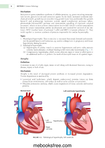
                           (chronic haemodynamic overload) leading to left ventricular hypertrophy (Fig. 1.3).
                         (b)  Compensatory hypertrophy, which occurs when an organ or tissue is called upon to
                           do additional work or to perform the work of destroyed tissue or of a paired organ.

                     Atrophy
                     Definition
                     A decrease in size of a body organ, tissue or cell along with decreased function, owing to
                     disease, injury or lack of use.

                     Mechanism
                     Atrophy  is  the  result  of  decreased  protein  synthesis  or  increased  protein  degradation.
                     Protein degradation is mediated by
                     •  Lysosomal  acid  hydrolases,  which  degrade  endocytosed  proteins  (taken  up  from
                       extracellular environment, cell surface as well as some cellular components).
                     •  Ubiquitin-proteasome pathway, which causes degradation of many cytosolic and nuclear
                       proteins.

                     Normal heart                               Left ventricular hypertrophy


                                               Left   Hypertrophy
                                               ventricle

                                               Right
                                               ventricle


                                  FIGURE 1.3.  Pathological hypertrophy, left ventricle.


                                  mebooksfree.com

