


Page 22 - Concise Pathology for Exam Preparation ( PDFDrive )
P. 22
1  Cell Injury and Cell Death  7


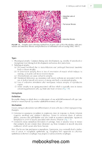
             FIGURE 1.4.  Atrophic testis showing marked loss of germ cells within the tubules, with peri-
             tubular and interstitial fibrosis and proliferation of interstitial cells of Leydig (H&E; 1003).


             Types
               1.  Physiological atrophy: Common during early development, eg, atrophy of notochord or
                thyroglossal duct during fetal development and uterus after parturition.
               2.  Pathological atrophy:
                 (a)  Decreased  workload  due  to  immobilization  and  prolonged  functional  inactivity
                   leads to disuse atrophy.
                 (b)  In denervation atrophy, there is loss of innervation of muscle which induces its
                   wasting, as in polio and motor neuron disease.
                 (c)  Atherosclerosis can cause ischaemic atrophy.
                  (d)  Nutritional deficiency, eg, marasmus and cancer cachexia are associated with the
                   use of skeletal muscle as a source of energy and lead to nutritional atrophy.
                 (e)  Loss of endocrine stimulation after menopause induces atrophy of reproductive
                   organs.
                 (f)  Senile atrophy is an ageing-associated cell loss which is typically seen in tissues
                   containing permanent cells, eg, brain and heart or testes (Fig. 1.4).

             Metaplasia
             Definition
             Reversible change in which there is replacement of one adult/differentiated cell type (epi-
             thelial or mesenchymal) by another adult/differentiated cell type.

             Mechanism
             Occurs owing to altered/aberrant differentiation of stem cells due to their reprogramming.
             Examples
             •  Columnar to squamous metaplasia in respiratory tract, in response to chronic irritation
               (cigarette  smoking)  and  vitamin-A  deficiency.  Stones  in  excretory  ducts  of  salivary
               glands, pancreas and gall bladder may also result in squamous metaplasia. Squamous
               metaplasia in cervix is usually associated with chronic infection (Fig. 1.5).
             •  Connective tissue metaplasia (formation of cartilage, bone or adipose tissue in tissues
               that normally do not contain these elements), eg, bone formation in muscle (myositis
               ossificans), which occurs after bone fracture.
             Note: The factors that predispose to metaplasia, if persistent, may eventually lead to induc-
             tion  of  cancer  in  metaplastic  epithelium,  eg,  metaplasia  from  squamous  to  columnar
             epithelium in Barrett’s oesophagus may progress to adenocarcinoma oesophagus.


                                  mebooksfree.com

