
Page 258 - Concise Pathology for Exam Preparation ( PDFDrive )
P. 258
10 Blood Vessels 243
Q. Describe the pathological features and clinical consequences of
atherosclerosis.
Ans. Early lesions show diffuse intimal thickening. Fatty streaks are the forerunners in the
evolution of atherosclerotic plaques.
1. Fatty streaks and dots
Salient features:
• Start by themselves and are harmless, but are considered earliest precursors of
atheromas.
• Usually begin in the first year of life, and are present in all children older than
10 years.
• Especially prominent in the aorta and other major arteries and are associated with
the known risk factors of atherosclerosis.
Gross: Multiple flat or slightly elevated, yellow intimal spots less than 1 mm in
diameter, which coalesce into elongated streaks, 1 cm or longer.
Microscopy: Composed of lipid-laden macrophages (foam cells) and a few T
lymphocytes.
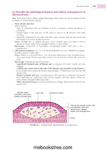
2. Atheromatous plaques (Fig. 10.3): Fully developed lesions, also called fibrous plaque,
fibrofatty plaque or atheroma
Gross: White to yellowish-white, 1–2 cm lesion raised above the luminal surface.
Has a grey-white fibrous cap and a central core of yellowish white soft, grumous
lipid.
Microscopy: Depends on age of the lesion
• Superficial (luminal) part of fibrous cap is composed of smooth muscle cells and
collagen.
• Cellular area under and to the side of the fibrous cap (shoulder of the lesion) is
more cellular and composed of foamy macrophages, T lymphocytes and few smooth
muscle cells.
• Deeper (central) soft core is located deep to the cap and is composed of extracel-
lular lipid material, cholesterol clefts (needle-shaped, cleft-like spaces), fibrin, ne-
crotic debris and lipid-laden foam cells.
• Older advanced lesions show dense hyalinized collagen, fibrous tissue and smooth
muscle cells.
Foam cells Cholesterol crystals
(cell debris, cholesterol
crystals, and foam cells)
Fibrous cap (smooth muscle cells,
macrophages, foam cells,
lymphocytes, collagen,
elastin, and proteoglycans)
Media
FIGURE.10.3. Diagrammatic representation of an atheroma.
mebooksfree.com