
Page 270 - Concise Pathology for Exam Preparation ( PDFDrive )
P. 270
11 Disorders of the Heart 255
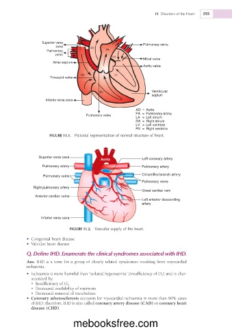
Superior vena Pulmonary veins
cava
Pulmonary
veins
Mitral valve
Atrial septum
Aortic valve
Tricuspid valve
Ventricular
septum
Inferior vena cava
AO = Aorta
PA = Pulmonary artery
Pulmonary valve
LA = Left atrium
RA = Right atrium
LV = Left ventricle
RV = Right ventricle
FIGURE 11.1. Pictorial representation of normal structure of heart.
Superior vena cava
Aorta Left coronary artery
Pulmonary artery Pulmonary artery
Pulmonary veins Circumflex branch artery
Pulmonary veins
Right pulmonary artery
Great cardiac vein
Anterior cardiac veins
Left anterior descending
artery
Inferior vena cava
FIGURE 11.2. Vascular supply of the heart.
• Congenital heart disease
• Valvular heart disease
Q. Define IHD. Enumerate the clinical syndromes associated with IHD.
Ans. IHD is a term for a group of closely related syndromes resulting from myocardial
ischaemia.
• Ischaemia is more harmful than ‘isolated hypoxaemia’ (insufficiency of O 2 ) and is char-
acterized by:
• Insufficiency of O 2
• Decreased availability of nutrients
• Decreased removal of metabolites
• Coronary atherosclerosis accounts for myocardial ischaemia in more than 90% cases
of IHD; therefore, IHD is also called coronary artery disease (CAD) or coronary heart
disease (CHD).
mebooksfree.com