
Page 275 - Concise Pathology for Exam Preparation ( PDFDrive )
P. 275
260 SECTION II Diseases of Organ Systems
• Morphological events
• ATP depletion leads to loss of contractility within a few minutes.
• A state of irreversible injury sets in within 20–40 min.
• Microvascular injury begins within 1 h.
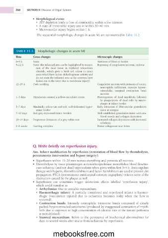
The sequential morphologic changes in acute MI are summarized in Table 11.2.
TABLE 11.2. Morphologic changes in acute MI
Time Gross changes Microscopic changes
0–4 h None Waviness of fibres at border
4–12 h None (the infarcted area can be highlighted by immer- Beginning of coagulation necrosis, oedema
sion of the dead tissue in triphenyl tetrazolium
chloride, which gives a brick red colour to intact
areas which have lactate dehydrogenase activity and
do not stain the infracted area as the enzymes have
leaked out in the latter due to membrane injury).
12–24 h Dark mottling Coagulative necrosis with pyknosis of nuclei;
neutrophilic infiltration, myocyte hypere-
osinophilia; marginal contraction band
necrosis
1–3 days Hyperaemia around a yellow-tan infarct centre Disintegration of dead myofibrils followed
by phagocytosis of dead cells by macro-
phages at infarct border
3–7 days Maximally yellow-tan and soft; well-delineated hyper- Early formation of fibrovascular granulation
aemic border tissue at margins
7–10 days Red-grey depressed infarct borders Well-established granulation tissue with new
blood vessels and collagen deposition
10–14 days Progressive formation of a grey-white scar Increased collagen deposition with decreased
cellularity
2–8 weeks Scarring complete Dense collagenous scar forms
Q. Write briefly on reperfusion injury.
Ans. Infarct modification by reperfusion (restoration of blood flow by thrombolysis,
percutaneous intervention and bypass surgery):
• Reperfusion within 15–20 min revives everything and prevents all necrosis.
• Thrombolysis by tissue plasminogen activator/streptokinase reestablishes blood flow/res-
cues ischaemic (and not dead) myocardium when given within first 3–4 h. Anticoagulant
therapy with heparin, thrombin inhibitors and factor Xa inhibitors are used to prevent clot
propagation. PTCA (percutaneous transluminal coronary angioplasty) relieves some of the
obstruction caused by the plaque as well.
• Reperfusion can sometimes trigger deleterious effects labelled ‘reperfusion injury’,
which could manifest as
• Arrhythmias: Due to unstable myocardium.
• Haemorrhagic infarct: A partially completed and reperfused infarct is haemor-
rhagic (vasculature injured due to ischaemia becomes leaky when the flow is
restored).
• Contraction bands: Intensely eosinophilic transverse bands composed of closely
packed hypercontracted sarcomeres (produced by exaggerated contraction of myofi-
brils due to exposure to high concentration of calcium ions at the instant perfusion
is reestablished).
• Stunned myocardium: Refers to the persistence of biochemical abnormalities for
days to several weeks after rescue from ischaemia by reperfusion.
mebooksfree.com