
Page 281 - Concise Pathology for Exam Preparation ( PDFDrive )
P. 281
266 SECTION II Diseases of Organ Systems
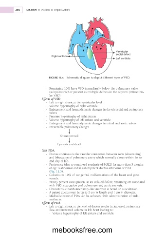
Ventricular
septal defect
Right ventricle
Left ventricle
FIGURE 11.4. Schematic diagram to depict different types of VSD.
- Remaining 10% have VSD immediately below the pulmonary valve
(subpulmonic) or present as multiple defects in the septum (infundibu-
lar VSD)
Effects of VSD
- Left to right shunt at the ventricular level
- Volume hypertrophy of right ventricle
- Enlargement and haemodynamic changes in the tricuspid and pulmonary
valves
- Pressure hypertrophy of right atrium
- Volume hypertrophy of left atrium and ventricle
- Enlargement and haemodynamic changes in mitral and aortic valves
- Irreversible pulmonary changes
Shunt reversal
Cyanosis and death
(iii) PDA
- Ductus arteriosus is the vascular connection between aorta (descending)
and bifurcation of pulmonary artery which normally closes within 1st to
2nd day of life.
- Persistence (due to continued synthesis of PGE2) for more than 3 months
of age is abnormal and is called patent ductus arteriosus or PDA
(Fig. 11.5).
- Constitutes 10% of congenital malformations of the heart and great
vessels.
- Ninety percent cases present as an isolated defect; remaining are associated
with VSD, coarctation and pulmonary and aortic stenosis.
- Characteristic harsh machinery-like murmur is heard on auscultation.
- A patent ductus may be up to 2 cm in length and 1 cm in diameter.
- Medical closure of PDA can be achieved with administration of indo-
methacin.
Effects of PDA
- Left to right shunt at the level of ductus results in increased pulmonary
flow and increased volume in left heart leading to:
- Volume hypertrophy of left atrium and ventricle
mebooksfree.com