
Page 276 - Concise Pathology for Exam Preparation ( PDFDrive )
P. 276
11 Disorders of the Heart 261
Q. Enumerate the types of myocardial infarcts. Differentiate between
transmural and subendocardial infarcts.
Ans. Depending on the thickness of the myocardium involved, myocardial infarcts are
classified into:
1. Transmural infarcts: Involve the whole thickness of the ventricular wall in the distribu-
tion of a single coronary artery
2. Subendocardial infarcts: Involve only the inner one-third to one-half of the ventricular
thickness
3. Multifocal microinfarcts: Multifocal microinfarction is seen when small intramural ves-
sels are involved by vasculitis, microembolization or vasospasm.
The differentiating features between transmural and subendocardial infarcts are enlisted
in Table 11.3.
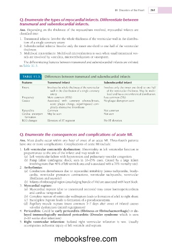
TABLE 11.3. Differences between transmural and subendocardial infarcts
Features Transmural infarct Subendocardial infarct
Extent Involves the whole thickness of the ventricular Involves only the inner one-third to one half
wall in the distribution of a single coronary of the ventricular thickness. May be multi-
artery focal and has a circumferential distribution.
Frequency More common (95%) Less common (5%)
Causes Associated with coronary atherosclerosis, No plaque disruption seen
acute plaque change, superimposed com-
pletely obstructive thrombosis
Epicarditis Common Not common
Cardiac aneurysm May be seen Not seen
formation
ECG changes Elevation of ST segment No ST elevation
Q. Enumerate the consequences and complications of acute MI.
Ans. Most deaths occur within one hour of onset of an acute MI. Three-fourth patients
have one or more complications. Complications of acute MI include:
1. Left ventricular contractile dysfunction: Abnormality in left ventricular function is
proportionate to the size of the infarct and may result in:
(a) Left ventricular failure with hypotension and pulmonary vascular congestion.
(b) Pump failure (cardiogenic shock; seen in 10–15% cases. Caused by a large infarct
involving more than 40% of left ventricle area and is associated with a 70% mortality rate).
2. Arrhythmias:
(a) Conduction disturbances due to myocardial irritability (sinus tachycardia, brady-
cardia, ventricular premature contractions, ventricular tachycardia, ventricular
fibrillation and asystole)
(b) Infarcts of inferoseptal region (area lodging bundle of His) are associated with heart block.
3. Myocardial rupture:
(a) Myocardial rupture (due to transmural necrosis) may cause haemopericardium
and cardiac tamponade
(b) Complete rupture of ventricular wall/septum leads to formation of a left to right shunt
(c) Incomplete rupture leads to formation of a pseudoaneurysm.
(d) Papillary muscle rupture (most common 3–7 days after onset of infarct) causes
valvular dysfunction (mitral regurgitation)
4. Pericarditis: Could be early pericarditis (fibrinous or fibrinohaemorrhagic) or de-
layed immunologically mediated pericarditis (Dressler syndrome which is seen
2–10 weeks after infarction)
5. Right ventricular infarction: Isolated right ventricular infarction is rare. Usually
accompanies ischaemic injury of left ventricle and septum
mebooksfree.com