
Page 282 - Concise Pathology for Exam Preparation ( PDFDrive )
P. 282
11 Disorders of the Heart 267
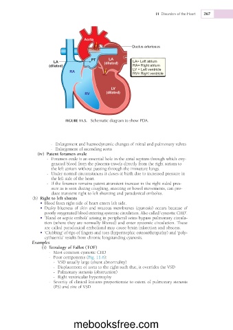
Aorta
Ductus arteriosus
PT LA
LA (dilated) LA= Left atrium
(dilated) RA= Right atrium
LV = Left ventricle
RA
RV= Right ventricle
LV
RV (dilated)
FIGURE 11.5. Schematic diagram to show PDA.
- Enlargement and haemodynamic changes of mitral and pulmonary valves
- Enlargement of ascending aorta
(iv) Patent foramen ovale
- Foramen ovale is an essential hole in the atrial septum through which oxy-
genated blood from the placenta travels directly from the right atrium to
the left atrium without passing through the immature lungs.
- Under normal circumstances it closes at birth due to increased pressure in
the left side of the heart.
- If the foramen remains patent atransient increase in the right sided pres-
sure as is seen during coughing, sneezing or bowel movements, can pro-
duce transient right to left shunting and paradoxical embolus.
(b) Right to left shunts
• Blood from right side of heart enters left side.
• Dusky blueness of skin and mucous membranes (cyanosis) occurs because of
poorly oxygenated blood entering systemic circulation. Also called ‘cyanotic CHD’.
• ‘Bland or septic emboli’ arising in peripheral veins bypass pulmonary circula-
tion (where they are normally filtered) and enter systemic circulation. These
are called paradoxical emboliand may cause brain infarction and abscess.
• ‘Clubbing’ of tips of fingers and toes (hypertrophic osteoarthropathy) and ‘poly-
cythaemia’ results from chronic longstanding cyanosis.
Examples
(i) Tetralogy of Fallot (TOF)
- Most common cyanotic CHD
- Four components (Fig. 11.6):
- VSD usually large (shunt abnormality)
- Displacement of aorta to the right such that, it overrides the VSD
- Pulmonary stenosis (obstruction)
- Right ventricular hypertrophy
- Severity of clinical features proportionate to extent of pulmonary stenosis
(PS) and size of VSD
mebooksfree.com