
Page 279 - Concise Pathology for Exam Preparation ( PDFDrive )
P. 279
264 SECTION II Diseases of Organ Systems
Genetic Factors
• Single gene mutations (genes encoding transcription factors required for normal cardiac
development, eg, GATA4, Tbx5 and genes encoding components of Notch pathway)
• Small chromosomal deletions, eg, deletion of chromosome 22q11.2 in DiGeorge syn-
drome, which leads to abnormal development of 4th bronchial arch and derivatives of
3rd and 4th pharyngeal pouches responsible for the development of heart.
• Additions and deletions of whole chromosomes, eg, trisomy of chromosomes13, 15, 18
and 21, and Turner syndrome
Maternal risks
Rubella, alcohol and drugs (teratogens)
Q. Classify congenital heart disease. Write briefly on the
clinicopathological features of the various types of CHD.
Ans. Classification and clinicopathological features of various types of CHD:
1. Malpositions of the heart (dextrocardia)
(a) Apex of the heart points to the right of the chest.
(b) May be accompanied by situs inversus; so, heart remains in normal position with
respect to the other organs.
(c) Isolated dextrocardia may be associated with major anomalies, eg, transposition of
aorta and transposition of great vessels.
2. Shunts: Abnormal communication between chambers or blood vessels
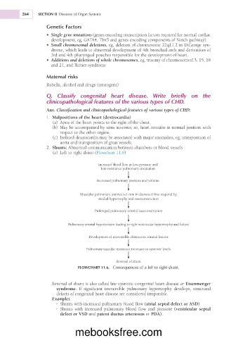
(a) Left to right shunt (Flowchart 11.6)
Increased blood flow in low-pressure and
low-resistance pulmonary circulation
Increased pulmonary pressure and volume
Muscular pulmonary arteries (<1 mm in diameter) first respond by
medial hypertrophy and vasoconstriction
Prolonged pulmonary arterial vasoconstriction
Pulmonary arterial hypertension leading to right ventricular hypertrophy and failure
Development of irreversible obstructive intimal lesions
Pulmonary vascular resistance increases to systemic levels
Reversal of shunt
FLOWCHART 11.6. Consequences of a left to right shunt.
Reversal of shunt is also called late cyanotic congenital heart disease or Eisenmenger
syndrome. If significant irreversible pulmonary hypertrophy develops, structural
defects of congenital heart disease are considered irreparable.
Examples
• Shunts with increased pulmonary blood flow (atrial septal defect or ASD)
• Shunts with increased pulmonary blood flow and pressure (ventricular septal
defect or VSD and patent ductus arteriosus or PDA).
mebooksfree.com